Fig. 1.
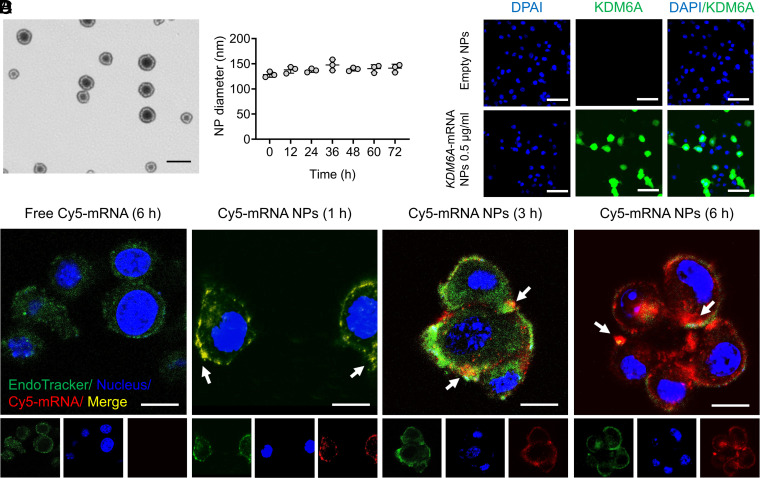
On-demand introduction of exogenous and functional proteins in Kdm6a-null BCa cells via mRNA NPs. (A) mRNA NP morphology was observed through TEM (Scale bar, 200 nm). (B) Stability of the mRNA NPs in PBS containing 10% serum throughout 72 h. The NP diameters were confirmed through dynamic light scattering measurements. (C) CLSM imaging of Kdm6a-null KU19-19 cells after different duration of incubation (1, 3, and 6 h) with Cy5-mRNA NPs (red). CellLight Late Endosomes-GFP was used to stain endosomes (green), and Hoechst 33342 was utilized to stain nuclei (blue). Cells after 6 h of incubation with free Cy5-mRNA were assigned to the control group (Scale bars, 10 µm). (D) IF staining of KDM6A in Kdm6a-null KU19-19 cells after treatment with empty NPs or KDM6A-mRNA NPs (Scale bars, 50 µm).

