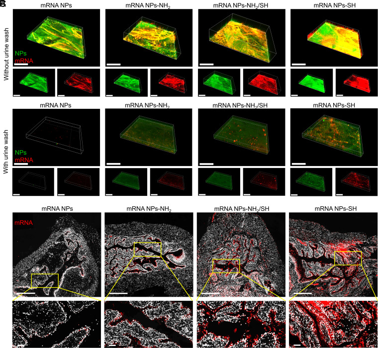
Fig. 4.
Engineering of surface mucoadhesive mRNA NPs for effective intravesical delivery of mRNA. (A) CLSM volume view images of mouse bladder walls after incubation (2 h) with nonmucoadhesive mRNA NPs, mucoadhesive mRNA NPs-NH2, mucoadhesive mRNA NPs-NH2/SH, or mucoadhesive mRNA NPs-SH. mRNA was labeled with Cy5 (red fluorescence), and NPs were labeled with Fluorescein (FITC) (green fluorescence) (Scale bar, 400 μm). (B) CLSM volume view images of mouse bladder walls after 2 h of incubation with different mRNA NPs, followed by another 3 h of incubation in urine (i.e., urine wash). mRNA was labeled with Cy5 (red fluorescence), and NPs were labeled with FITC (green fluorescence) (Scale bar, 400 μm). (C) Sections of the bladder tissues from mice treating with nonmucoadhesive mRNA NPs, mucoadhesive mRNA NPs-NH2, mucoadhesive mRNA NPs-NH2/SH, or mucoadhesive mRNA NPs-SH via intravesical administration (Scale bars in raw images, 500 µm; Scale bars in enlarged images, 50 µm). mRNA was labeled with Cy5 (red fluorescence).

