Fig. 8.
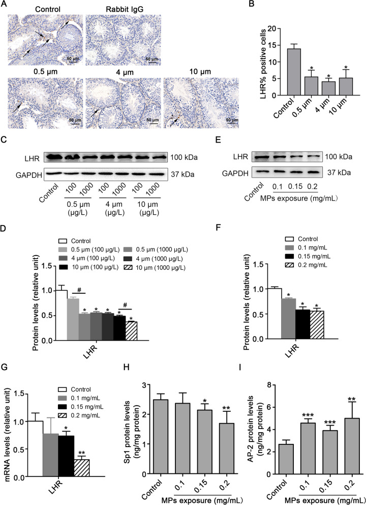
PS-MPs induced a decrease in testosterone levels by reducing LHR levels. A, B Mice were given drinking water containing different sizes of PS-MPs for 180 continuous days. The testis tissue sections were prepared for immunohistochemical staining with LHR antibody, arrows: positive expression (scale bar = 50 µm, n = 3). Percent of positivity was calculated based on the percentage of LHR positive cells out of the total number of cells in an image (*P < 0.05 vs. the control). C, D Mice were given drinking water containing various sizes of PS-MPs for 180 continuous days. The expression of LHR in testes was measured by western blotting. The expression levels were quantified with ImageJ and expressed as means ± SD (n = 3; *P < 0.05 vs. control; #P < 0.05 vs. 100 μg/L group). E, F Primary Leydig cells were exposed to 0.1, 0.15, and 0.2 mg/mL PS-MPs with a diameter of 0.5 μm for 24 h. The expression of LHR in cells was analyzed by western blotting. The expression levels were quantified with ImageJ and expressed as means ± SD (n = 3; *P < 0.05 vs. control). G The mRNA expression levels of LHR in Leydig cells after treatment with PS-MPs were determined by qRT-PCR (n = 3; *P < 0.05, **P < 0.01 vs. control). (H, I) The contents of Sp1 and AP-2 were detected by ELISA (n = 3; *P < 0.05, **P < 0.01, ***P < 0.001 vs. control)

