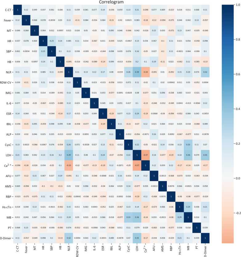
Fig. 2.
The heat map of correlation between features. Color indicates the value of the correlation coefficient (r). The color intensity is proportional to the correlation coefficient (r), with positive correlations (r > 0) shown and negative correlations (r < 0), lower intensive color indicates lower correlations, in the study, 23 features selected by machine learning techniques, each feature is weakly correlated with each other (r < 0.4). Twenty-three features including C-CT, fever, MT, HR, SBP, HB, NLR, RDW-CV, IMG, IL-6, ESR, IBIL, ALP CysC, LDH, Ca2+, AFU, AMS, RBP, Hs-cTn, MB, PT(s), D-dimer. C-CT chest computed tomography, MT malignant tumor, HR heart rate, SBP systolic blood pressure, NLR neutrophil-to-lymphocyte ratio, HB hemoglobin concentration, RDW-CV red cell volume distribution width, LDH lactate dehydrogenase, IBIL indirect bilirubin, PT prothrombin time, ESR erythrocyte sedimentation rate, AFU α-fucosidase, RBP retinol-Binding protein, IL-6 interleukin-6, Hs-cTn hypersensitive troponin, AMS amylase, CysC cystatin C, IMG immature granulocyte, ALP alkaline phosphatas, MB myoglobin

