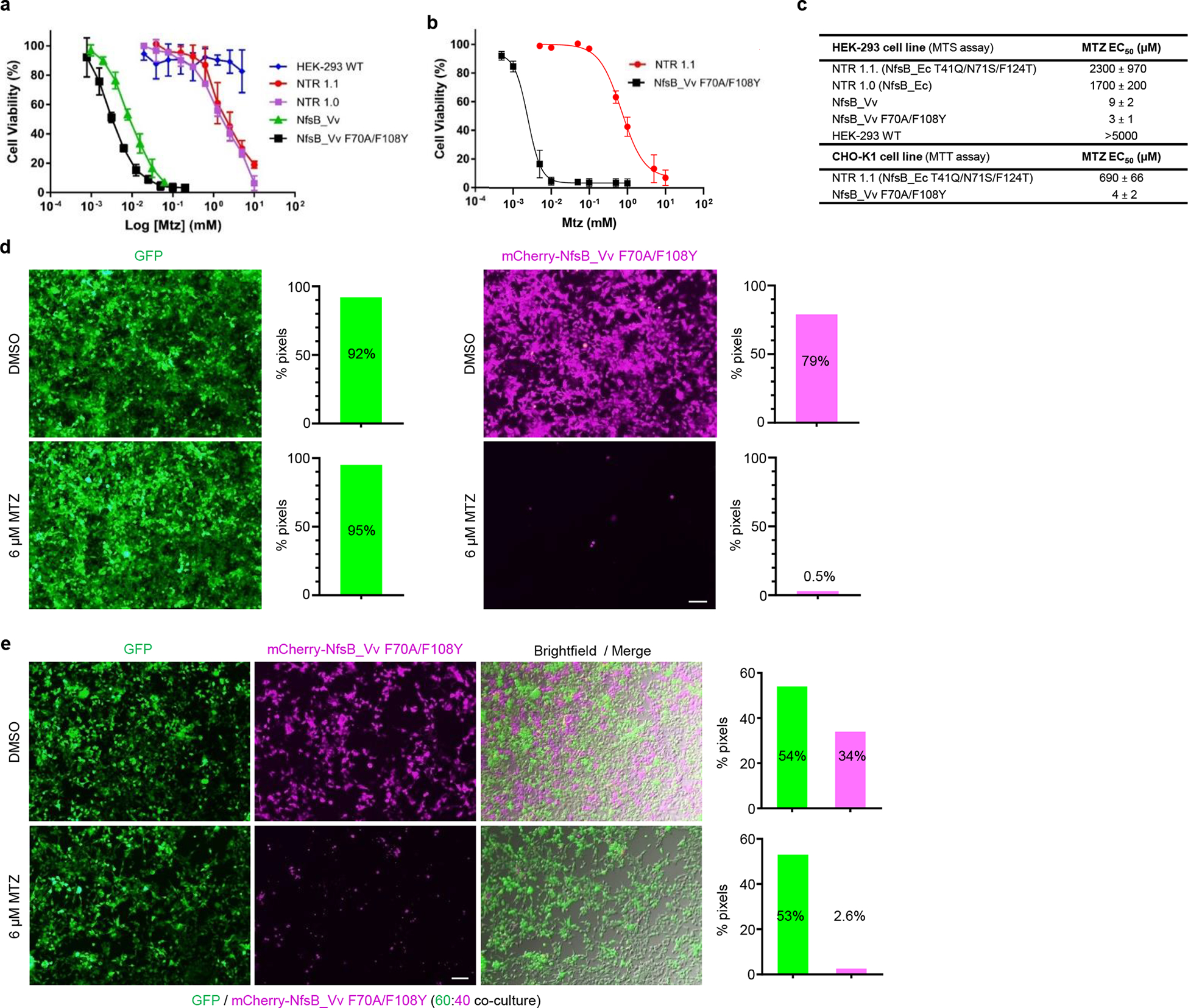
Fig. 2: Targeted ablation of mammalian cells is enhanced with NsfB_Vv F70A/F108Y.

a-b, MTZ dose-response cell viability assays of NTR variants in mammalian cells tested across the indicated MTZ concentrations. a, NTR variants stably expressed in HEK-293, n=5 biologically independent experiments for all cell lines other than wild-type HEK-293 cells (n=4) and those expressing NsfB_Vv F70A/F108Y (n=3) or NfsB_Vv (n=9). b, NTR variants stably expressed in CHO-K1 cells, n=3 biologically independent experiments. Survival rates were measured using MTS (a) or MTT (b) assays and data presented are means ± SD. c, MTZ EC50 values for mammalian HEK-293 and CHO-K1 cell lines stably over-expressing the indicated NTR enzyme variants. Data presented are means ± SD. d-e, Images and quantification of MTZ-induced ablation of transgenic HEK-293 cell lines. Cells expressing GFP or co-expressing mCherry and NfsB_Vv F70A/F108Y were cultured in isolation (d), or as a 60:40 co-culture of both cell lines (e). All cells were treated with 0.01% DMSO or 6 μM MTZ for 48 h and cell viability was assessed qualitatively by fluorescence microscopy and quantitatively by pixel counts (fluorescent pixels/total pixels), n=3 biologically independent experiments per condition. Merged brightfield and fluorescence images (e) confirm loss of NfsB_Vv F70A/F108Y expressing cells as opposed to loss of reporter expression. Scale bars = 100 microns.
