
Figure 7. Silencing VSCTs in vivo in freely-moving adult mice perturbs locomotor ability.
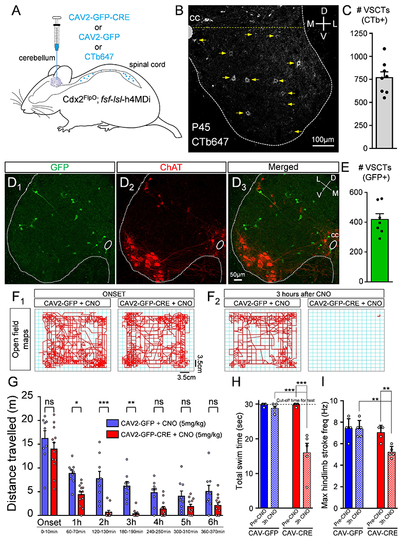
(A) CAV2-GFP-CRE or CAV2-GFP or CTb647 (for counting) was injected in cerebellum at ~P21 in Cdx2Flpo::fsf-lsl-h4MDi mice. (B) CTb647+ neurons in the L1 segment in a P45 mouse. Neurons ventral to the yellow dotted line were defined as VSCTs. (C) Total number of VSCTs labelled with CTb647 in the L1 segment (N=8). GFP in the L1 segment (D1: green), ChAT (D2: red) and merged image (D3). (E) Total number of GFP+ VSCTs in mice injected with CAV2-GFP-CRE (N=7). Maps of distance travelled by a mouse in open field assay in 10mins at the onset (F1) and after 3hours (F2) for each group (age: P45). (G) Distance travelled by mice in bins of 10mins duration for each hour after CNO injection (N=9 CAV2-GFP mice, blue; and N=12 CAV2-GFP-CRE mice, red). Each point represents a single mouse, ns: no significance (p>0.05), * p<0.05, *** p<0.001, One WayANOVA, Tukey’s post hoc test. (H) Total swim time for control (CAV2-GFP; blue) and CAV2-CRE (red) mice, before (Pre-CNO) and 3 hours after 10mg/kg CNO (3h CNO). (I) Maximum hindlimb stroke frequency during periods of swimming for the two groups. ** p<0.01, *** p<0.001, One Way ANOVA, Tukey’s post hoc test for both H and I. Data are represented as mean ± SEM. See also Figs. S8 and S9 and Videos 1, 2 and 3.