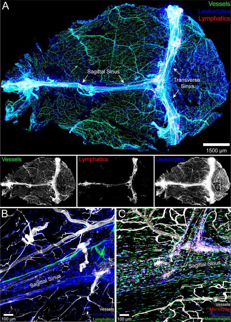
Figure 2. Anatomy of the meninges during steady state.

A.) The naïve meninges beneath the skull bone of an 8-week-old C57BL/6J mouse were harvested and imaged in 3D using confocal microscopy. The whole mount consisting primarily of dura and arachnoid mater shows the distribution of blood vessels labeled intravenously with fluorescent tomato lectin (green), CD45+ leukocytes (blue), and Lyve1+ lymphatic vessels (red). The individual grayscale images for each channel are shown beneath the 3-color overlay. The sagittal and transverse sinuses are also labeled. Note that the meningeal lymphatics are juxtaposed to the dural venous sinuses. B.) A three color zoomed confocal image from a meningeal whole mount shows a Lyve1+ lymphatic vessel adjacent to the superior sagittal sinus. CD45+ leukocytes are shown in blue and tomato lectin+ blood vessels in white. C.) A confocal image captured from a meningeal whole mount shows the distribution of CCR2rfp/+ monocytes (red) and CX3CR1gfp/+ meningeal macrophages (green) along the superior sagittal sinus. Blood vessels are shown in white.
