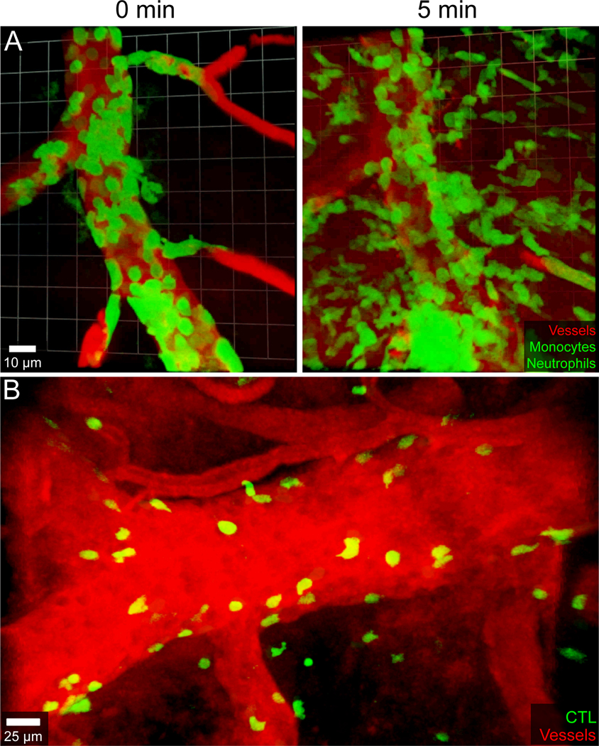
Figure 4. Immune mediated damage to CNS vasculature during viral meningitis and cerebral malaria.
A.) An intravital two-photon time lapse captured through the thinned skull window of a LysMgfp/+ mouse six days following intracerebral inoculation of LCMV Armstrong shows myelomonocytic cells (green) synchronously extravasating from a meningeal blood vessel. Quantum dots (red) were injected intravenously to visualize blood vessels. Note the massive extravasation of myelomonocytic cells at 5 min, which coincides with leakage of quantum dots into the extravascular space. B.) Parasite-specific cytotoxic lymphocytes (CTL, green) tagged with a fluorescent protein were visualized at the peak of disease (day 6) in a C57BL/6J mouse infected intravenously with 106 red blood cells parasitized by Plasmodium berghei ANKA. CTL were observed attacking meningeal and cerebral vasculature, arresting along both the luminal and abluminal surfaces. These CTL engagements are responsible for fatal vascular breakdown during cerebral malaria in both mice and humans. Blood vessels were visualized with Evans Blue (red).

