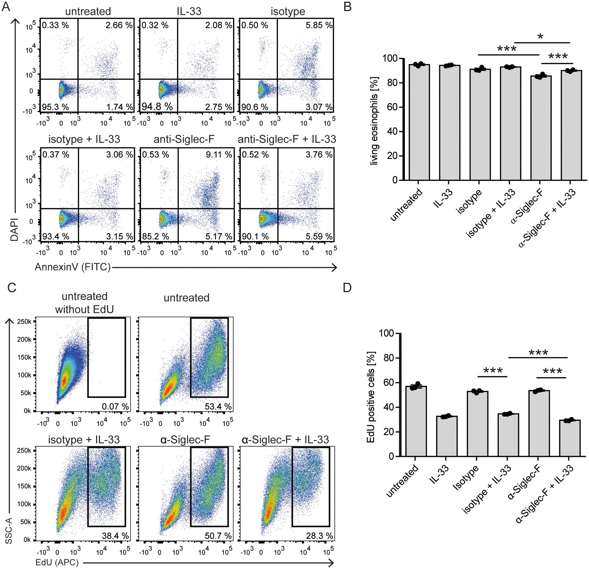
Figure 2: Siglec-F signaling barely affects survival and proliferation of IL-33-stimulated eosinophils.

A, B) Survival analysis. BMDE of culture d14 were stimulated with anti-Siglec-F or isotype control and optional IL-33 for 24 hrs and stained with AnnexinV and DAPI. Cells were pregated on eosinophils defined as CCR3+ cells (as in Suppl. Fig. 1B). A) Representative flow cytometry plots and B) Quantification of AnnexinV and DAPI double negative eosinophils. Shown are technical triplicates from one representative BMDE culture representative for three biologically distinct cultures. C, D) Proliferation analysis. BMDEs of culture d8 were stimulated with indicated conditions for 48 hrs and EdU was added for the last 24 hrs. C) Representative flow cytometry plots for incorporated EdU pregated on eosinophils and expected eosinophil precursors based on Siglec-F and CCR3 expression as in Suppl. Fig 1B. D) Quantification of the percentage of EdU+ cells from technical triplicates of one BMDE culture representative for two biologically distinct cultures. For B) and D) Mean ± SEM is shown. Two-way ANOVA with Bonferroni posttests. Only selected significances that highlight IL-33 and Siglec-F mediated effects are indicated; *p <0.05, ***p <0.001.
