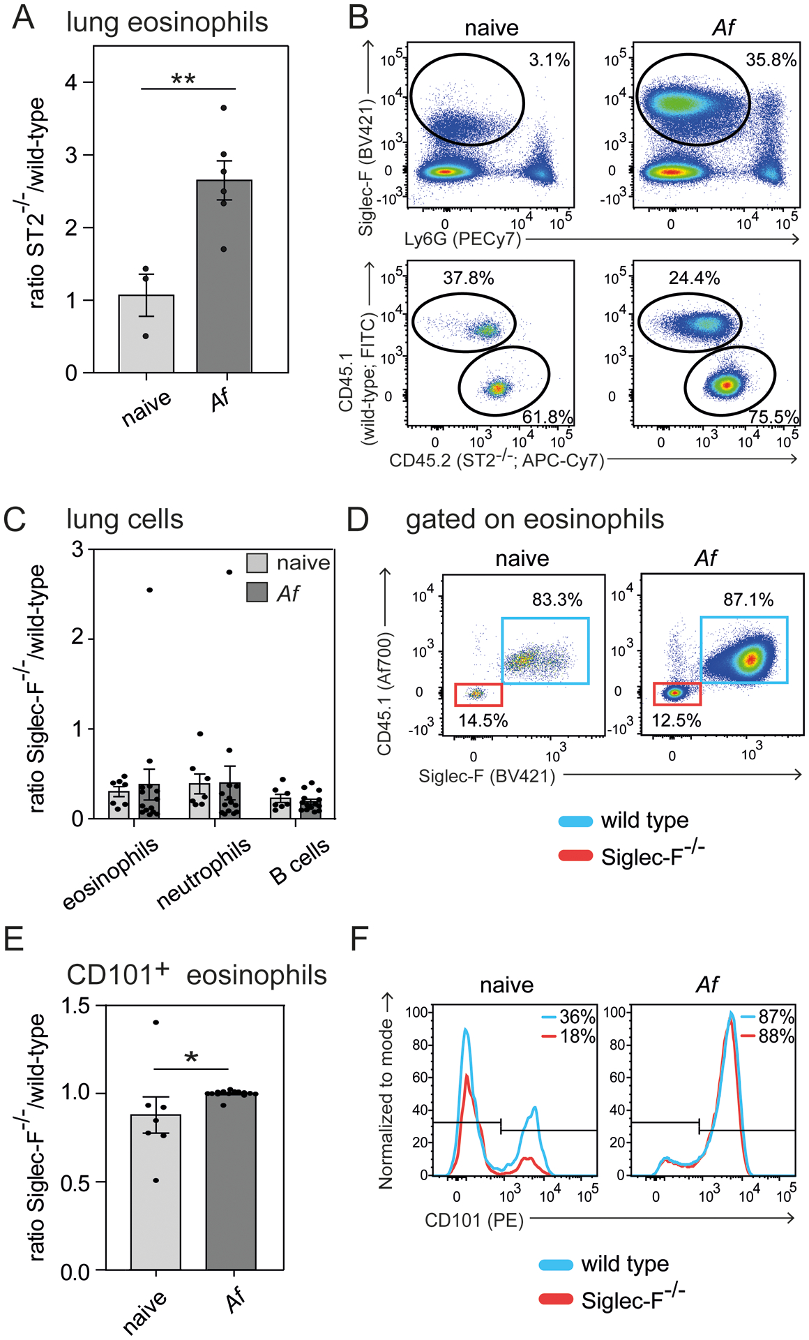
Figure 3: IL-33- rather than Siglec-F signaling dampens eosinophil expansion in A. fumigatus-elicited lung inflammation.

Competitive mixed bone marrow chimeras for ST2−/− (A and B) or Siglec-F−/− (C-F) (both CD45.2) generated in a 50:50 mix with CD45.1 congenic wild-type bone marrow were subjected to intranasal A. fumigatus (Af) infection and analyzed on day 17 by flow cytometry. A) Ratio of ST2−/− knock-out against wild-type eosinophils of the lung. B) Representative flow cytometric plots highlight eosinophil gating (Siglec-F+Ly6G−) and further subgating of eosinophils based on congenic markers. C) Ratio of Siglec-F−/− against wild-type cells for indicated populations. D) Representative flow cytometric plots display pre-gated eosinophils (Suppl. Fig. 1C) subdivided into Siglec-F−/− (Siglec-F−CD45.1−, red) or wild-type derived eosinophils (Siglec-F+CD45.1+, blue). E) Ratio of Siglec-F−/− against wild-type CD101+ eosinophils in the lung. F) Representative histograms for CD101 expression on eosinophils. n=2 independent experiments with a total of 3–6 mice (A and B) or 7–14 mice (C-F) per group. Two-way-ANOVA with Bonferroni posttests was performed to determine significance for C). Students t-test was performed for eosinophil ratio in ST2−/− chimeras (A) and frequency of CD101+ eosinophils (E). Bars show the mean ± SEM; *p < 0.05; **p < 0.01.
