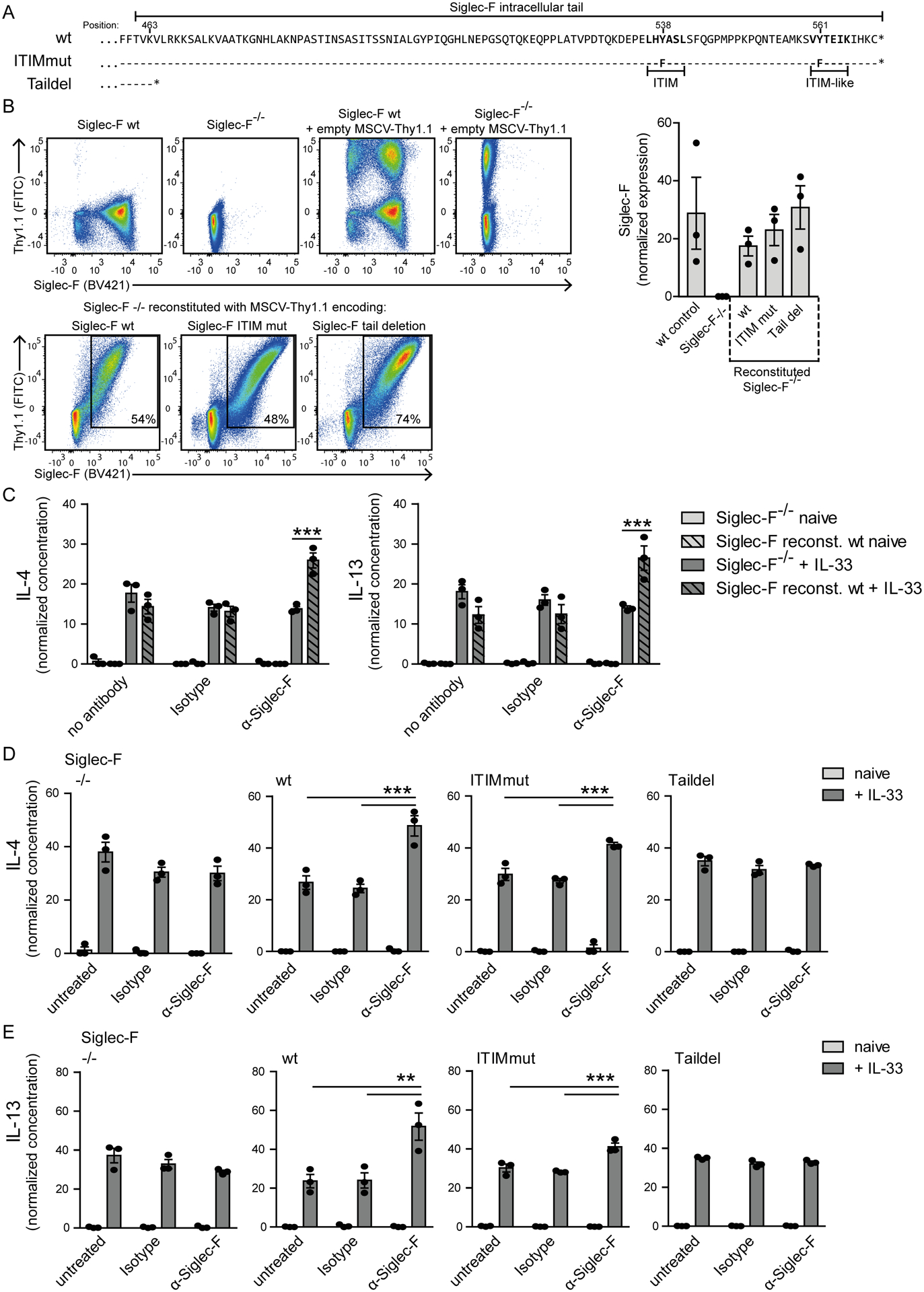
Figure 8: The cytoplasmic tail of Siglec-F promotes the enhanced cytokine/chemokine secretion from IL-33-stimulated eosinophils but the ITIM motifs are dispensable.

Siglec-F−/− bone marrow cells were transduced with Siglec-F variants-encoding MSCV-IRES-Thy1.1 retroviral vectors. A) Illustration of cytoplasmic tail sequences for Siglec-F variants. Dashes indicate matched amino acids and stars position of stop codons. B) Surface expression of Siglec-F and Thy1.1 from indicated populations. The bar graph shows the mean Siglec-F expression levels between wild-type BMDE cultures and retrovirally expressed Siglec-F variants. C) IL-4 and IL-13 concentrations in supernatants for MSCV-IRES-Thy1.1 transduced Siglec-F−/− and wild-type reconstituted Siglec-F−/− BMDE. Cells were unstimulated (light grey) or IL-33-stimulated (dark grey) and optionally treated with anti-Siglec-F or isotype antibody. D) and E) IL-4 (D) and IL-13 (E) normalized concentrations in supernatants of BMDE reconstituted with indicated Siglec-F mutants upon indicated treatment. In D) and E) culture conditions are compared individually for each variant. Normalization between experiments was performed with sum of replicate normalization. Bars show the mean ± SEM from n = 3 different cultures. One-way ANOVA with Bonferroni posttests was performed to determine significance. Only selected significances that highlight IL-33 and Siglec-F mediated effector release are indicated; *p < 0.05; **p < 0.01; ***p < 0.001.
