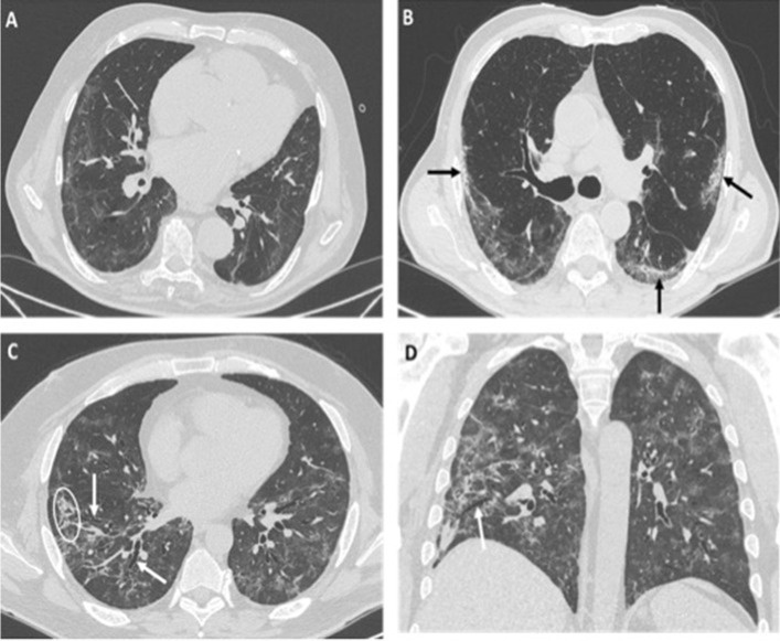
Fig. 1.
a–d HRCT images of different patients with COVID-19 pneumonia showing typical imaging features indicative of fibrosis: irregular interface (a), sub-pleural bandlike parenchymal consolidations (black arrows in b), coarse reticular opacities (white circle in c) and bronchial dilatation, observed both on axial image (white arrows in c) and on coronal reconstruction (white arrow in d)

