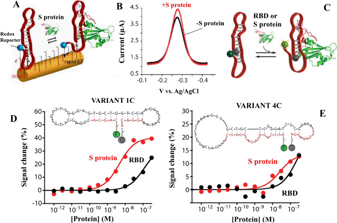
Fig. 10.
A Mechanism of electron exchanging in the interrogating electrode. B Single-step detection of S-protein by DPV. C Scheme of fluorophore–quencher couple at the two ends of the aptamer for studying binding activities by fluorescence spectroscopy. D and E Binding activities of the 1C and 4C variants of aptamers labelled with a fluorophore–quencher couple at the two ends against the SARS-CoV-2 RBD (black curves) and the S protein (red curves) in solution. Reproduced with permission from [122]

