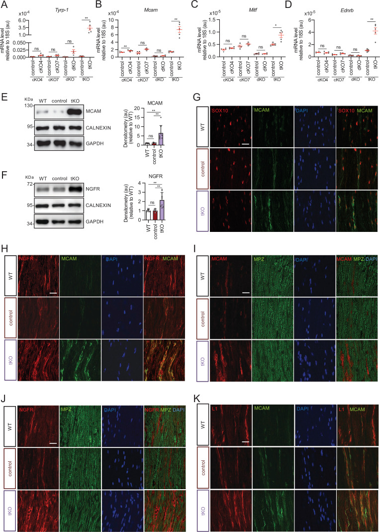
Figure 3. Melanocyte lineage markers are expressed by nonmyelinating Schwann cells of the Remak bundles in the sciatic nerves of the tKO.
(A) mRNA for Tyrp1 is dramatically increased by 1.081-fold in the tKO (3.35 ± 0.71 × 10⁻⁴ au in the tKO versus 0.11 ± 0.05 × 10⁻⁶ au in controls; p = 0.0092) whereas no changes were found in the cKO4, cKO7 neither dKO sciatic nerves. (B) mRNA for Mcam is also upregulated (5.13-fold) in the tKO (7.39 ± 0.79 × 10⁻⁴ au in the tKO versus 1.44 ± 0.06 × 10⁻⁴ au in controls) with only minor o no changes at all for the other genotypes. The same although less marked (1.74-fold) for Mitf (0.87 ± 0.09 × 10⁻⁵ au in the tKO versus 0.50 ± 0.02 × 10⁻⁵ au in controls; p = 0.0128) (C) and Ednrb (4.1-fold; 4.23 ± 0.52 × 10⁻⁵ au in the tKO versus 1.04 ± 0.09 × 10⁻⁵ au in controls; p = 0.0032) (D). RT-qPCR with mouse-specific primers for the indicated genes was performed. Graph shows a scatter plot for the ΔCt (which include also the mean ± standard error [SE]) of the gene normalized to the housekeeping 18S. Four to five mice per genotype were used. Data were analyzed with the unpaired t-test with Welch’s correlation. (E) MCAM protein levels in the sciatic nerves of the tKO. A representative Western blot of protein extracts from wild-type (C57BL/6), control and tKO sciatic nerves is shown. MCAM protein was increased by 7.6-fold in the tKO (9.93 ± 1.75 au in the tKO versus 1.30 ± 0.13 in controls; p = 0.0003). (F) NGFR protein was increased by 2.15-fold (2.16 ± 0.29 in the tKO versus 1.005 ± 0.09 in controls; p = 0.0003). Four to eight WB of the same number of animals per genotype were quantified. Data were analyzed with the one-way analysis of variance (ANOVA) Tukey’s test. (G) MCAM signal colocalizes with SOX10. (H) MCAM signal colocalizes with NGFR. (I) MCAM is not expressed by myelin-forming Schwann cells (MPZ+). (J) Same happens with NGFR. (K) MCAM signal colocalizes with L1cam, a marker of the nonmyelin-forming Schwann cells of the Remak bundles. P60 sciatic nerves were fixed and submitted to immunofluorescence with the indicated antibodies. Nuclei were counterstained with Hoechst. Representative confocal images of sections obtained from the sciatic nerves of wild-type (WT), control, and tKO mice are shown. Scale bar: 20 μm (*p < 0.05; **p < 0.01; ***p < 0.001; ns: no significant). See source data file one online (graphs source data) for more details.

