FIG 3.
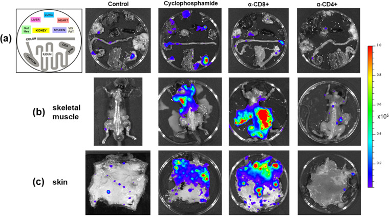
Tissue-specific impact of T cell depletion on parasite burden. C3H/HeN mice chronically infected with T. cruzi CL Luc::mNeon were treated with cyclophosphamide, anti-CD4, or anti-CD8 antibodies as outlined in the legend to Fig. 1. Sixteen days after treatment initiation, organs and tissues were examined by ex vivo imaging (52) (Materials and Methods). (a) Representative bioluminescence images of internal organs from treated mice arranged as shown on the left. Mes, mesentery; Vis, visceral (fat); OES, oesophagus; STM, stomach. (b) Dorsal bioluminescence images following removal of internal organs, fur, skin, and major adipose depots (Materials and Methods). (c) Ex vivo bioluminescence imaging of skin (adipose tissue removed). Radiance (photons/s/cm2/sr) is on a linear-scale pseudocolor heat map. The heat map image of skeletal bioluminescence after treatment with anti-CD8 antibodies is shown at an increased minimum and maximum radiance (1 × 10⁴ to 1 × 10⁶) to avoid saturation of the image. The complete radiance data set is shown in Fig. 2.

