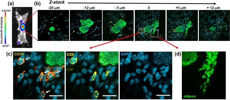
FIG 8.
Incomplete recruitment of leukocytes to parasite nests allows progression of T. cruzi through the full intracellular infection cycle. (a) An intense bioluminescent focus in a chronic stage distal colon viewed by ex vivo imaging (Materials and Methods). Radiance (photons/s/cm2/sr) is shown on a linear-scale pseudocolor heat map. (b) Confocal imaging of the corresponding parasite nest showing representative serial Z-stack images taken along the depth of the infected cell. The z axis position relative to the center of the nest is indicated above each of the images. Parasite numbers (>1,000) were established from green fluorescence and the characteristic DAPI staining of the parasite kinetoplast DNA (the mitochondrial genome) (18) (blue). Infiltrating leukocytes (orange) were identified by staining with anti-CD45 antibodies (Materials and Methods). Bar = 20 μm. (c) Enlarged images of a small cluster of infiltrating CD45+ (orange) and CD8+ (yellow) cells in close vicinity to the nest. White arrows indicate leukocytes corresponding to CD8+ T cells. (d) Egress of differentiated trypomastigotes into the extracellular environment. Data from the infected cell captured in these images were not included in Fig. 7, since the parasite burden was too great to determine numbers with precision.

