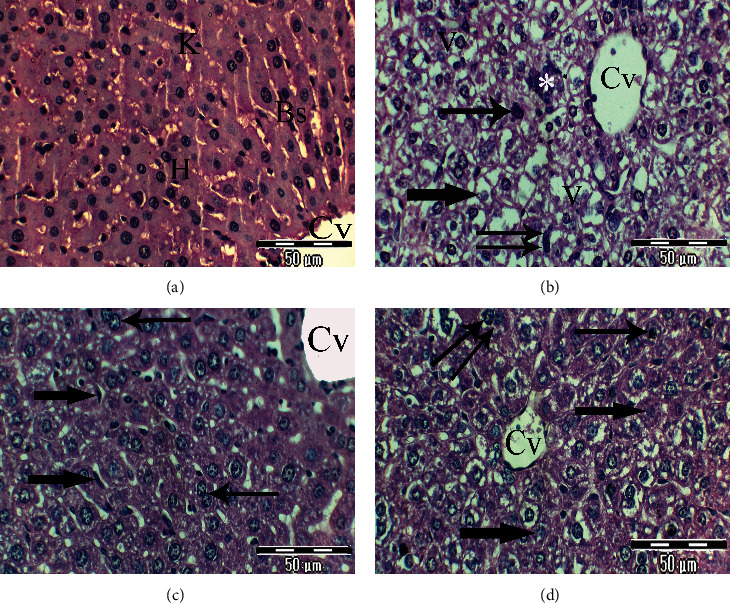
Figure 2.

(a–d) Photomicrographs of rat liver sections of different experimental groups stained with Haematoxylin & Eosin. (a) Liver section of control rats showing normal hexagonal hepatic lobules; the central vein (Cv) is located in the central part. Hepatocytes (H) are organized into hepatic cords and separated by normal Kupffer (K) cells in blood sinusoids (Bs) (×400). (b) Liver section of rats from the untreated lead-intoxicated group showing a noticeable disorganized liver section, degenerative hepatocytes with a vacuolated cytoplasm (V) and demarcated membrane, dilated and widening central vein (Cv), noticeable cellular infiltration (∗), pronounced nuclear changes such as pyknotic nuclei (thin arrows) and karyolitic ones (thick arrows), and distinct phagocytic Kupffer cells (double arrows) (×400). (c) Liver section of rats from the CRB-administered group exhibits noticeable improvement of hepatic architecture, hepatic cords radiating from the normal central vein (Cv), normal appearance of most nuclei but increase in the number of binucleated hepatocytes (thin arrows), and regular blood sinusoid network with Kupffer cell activity (thick arrows) (×400). (d) Section of the rat liver from the HRB-administered group shows mild improvement of hepatic tissue, most of hepatocytes are intact, some had a degenerative and vacuolated cytoplasm, most of the nuclei are normal, and others are pyknotic (thin arrows), karyolitic (thick arrows), and megakaryocytic ones (double arrows) (×400).
