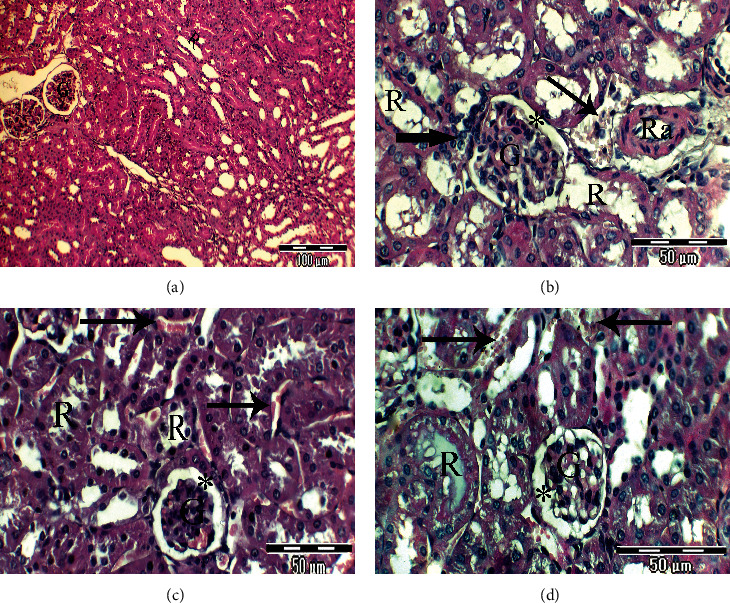
Figure 3.

(a–d) Photomicrographs of rat kidney sections of different experimental groups stained with Haematoxylin & Eosin. (a) Renal cortex of rats in the control group has a normal body structure of renal glomeruli (G) and renal tubules (×200). (b) High-magnification view of a portion of the rat kidney from the untreated lead-intoxicated group exhibiting disorganized kidney anatomy, irregular glomeruli with irregular and atrophied mesangial areas (star), necrosis of renal tubules with atrophy and destruction of their lining epithelium (thick arrow), severe congestion of the renal vein, and fibrinoid necrosis of the renal artery (Ra) (×400). (c) High-magnification view of a portion of the rat kidney from the CRB-administered group exhibits the normal anatomy of most glomeruli and a mesangial section (star) that appears normal, elongated and distended renal tubules, and intertubular hemorrhage (arrows) (×400). (d) High-magnification view of a portion of the rat kidney from the HRB-administered group showing mild improvement of the kidney anatomy, glomeruli with an irregular mesangial area (star), atrophy of some renal tubules, others with hyaline casts, and intertubular hemorrhage (thick arrows) (×400).
