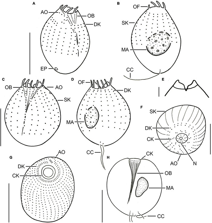
FIGURE 2.

Different urotrich morphotypes after protargol staining. (A,B) Ventral and dorsal views of a specimen of strain CIL-2017/24 from Lake Mondsee tentatively identified as Urotricha agilis. Occasionally, only the ciliated basal bodies of the adoral organelles stain. (C–F) Urotricha furcata, specimens of strain CIL-2019/6 from Lake Zurich. Right (C) and left (D) lateral views of same specimen. Optical section of the protruding oral region (E). Oblique top view (F). (G,H) Specimens of strain CIL-2019/1 from Lake Zurich tentatively identified as U. castalia. Oblique top view (G); the concentric circles denote the circumoral kinety. Schematic optical section (H). AO, adoral organelles; CC, caudal cilia; CK, circumoral kinety; DK, dikinetids at anterior end of somatic kineties; EP, excretory pore; MA, macronucleus; N, nematodesmata (oral basket rods); OB, oral basket; OF, oral flaps; SK, somatic kineties. Scale bars 10 μm (A–D,F), 2 μm (E), and 20 μm (G,H).
