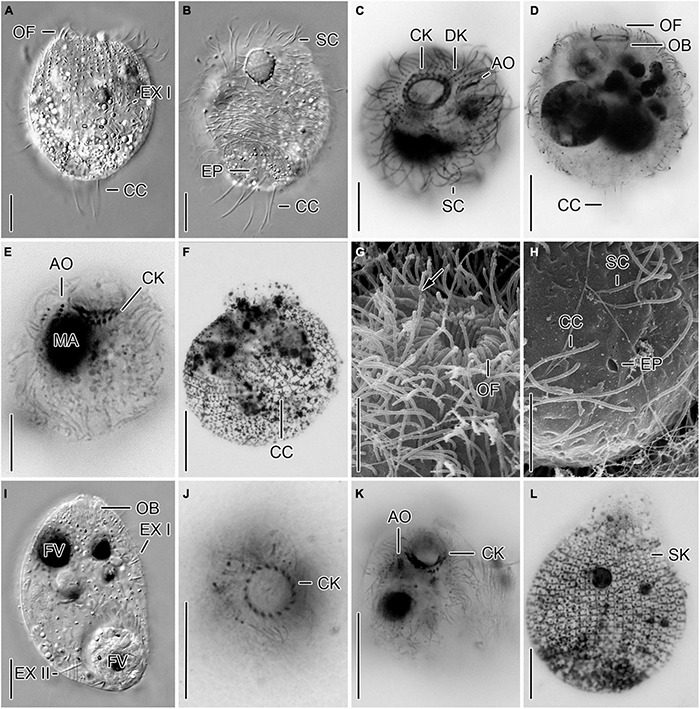
FIGURE 5.

Cryptic species tentatively identified as Urotricha castalia. One species (A–H) is represented by specimens of strains CIL-2019/2 (A,B), CIL-2019/1 (C,D,F–H), and of the non-clonal original culture (E) from Lake Zurich, the other species (I–L) by specimens of strains CIL-2017/27 (I) and CIL-2017/25 (J–L) from Lake Mondsee in vivo (A,B,I), after protargol staining (C–E,J,K), after dry silver nitrate staining (F,L), and in the scanning electron microscope (G,H). (A,E,L) Lateral views. Occasionally, only the ciliated basal bodies of the adoral organelles stain (E). The silverline pattern is typical (L). (B) Ventral view. The excretory pore is within the circle of caudal cilia. (C,G,K) Oblique top views. Arrow denotes distinct ridges between adoral organelles. (D,I) Longitudinal optical sections. (F,H) Oblique posterior polar views. The silverline pattern is composed of polygonal meshes in the unciliated cell portion and the caudal cilia insert in rather circular meshes (F). The excretory pore is within the circle of caudal cilia (H). (J) Top view. AO, adoral organelles; CC, caudal cilia; CK, circumoral kinety; DK, dikinetids at anterior end of somatic kineties; EP, excretory pore; EX I, small extrusomes in ciliated anterior cell portion; EX II, large extrusomes in unciliated posterior cell portion; FV, food vacuole; MA, macronucleus; OB, oral basket; OF, oral flaps; SC, somatic cilia; SK, somatic kineties. Scale bars 10 μm (A–F,I–K) and 5 μm (G,H,L).
