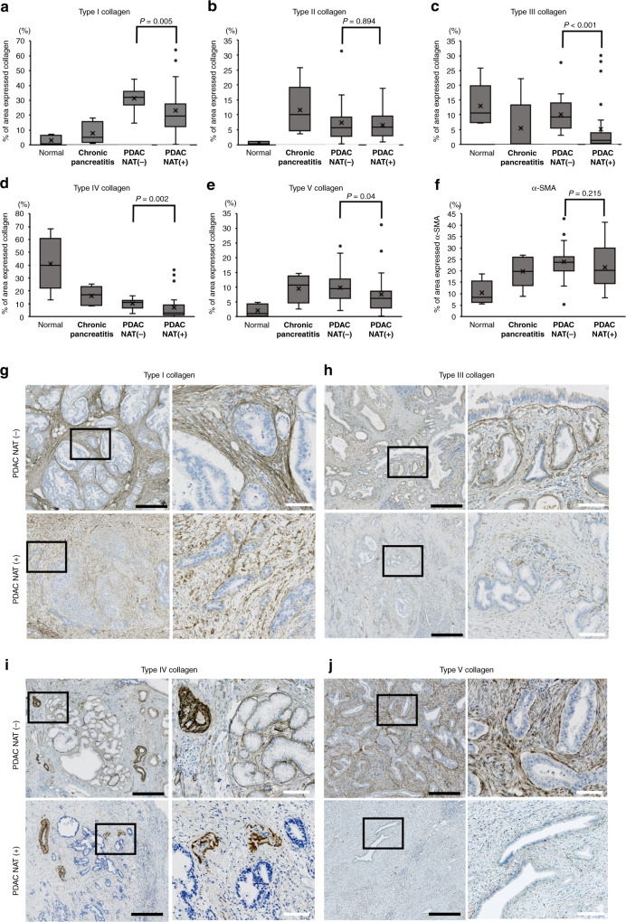
Fig. 1. Comparison of the collagen-expressed area in the four pancreatic conditions.
a–f The area occupied by the collagens and alpha-smooth muscle actin (α-SMA) was quantified using an automatic image analyzer after immunohistochemistry. Four pancreatic conditions were compared; normal pancreas (n = 5), chronic pancreatitis (n = 5), pancreatic cancer (PDAC) without neoadjuvant therapy (NAT) (n = 20), and PDAC with effective NAT (n = 25). Mann–Whitney U test was used for statistical analysis. g–j Immunohistochemical findings of collagens in PDAC tissues with or without NAT. For comparison, a pair of PDAC cases with effective NAT or without NAT is displayed. The left picture shows the low-power view, and the right picture shows the high-power view corresponding to the field of black square in the left picture. Black bars = 500 µm. White bars = 100 µm.

