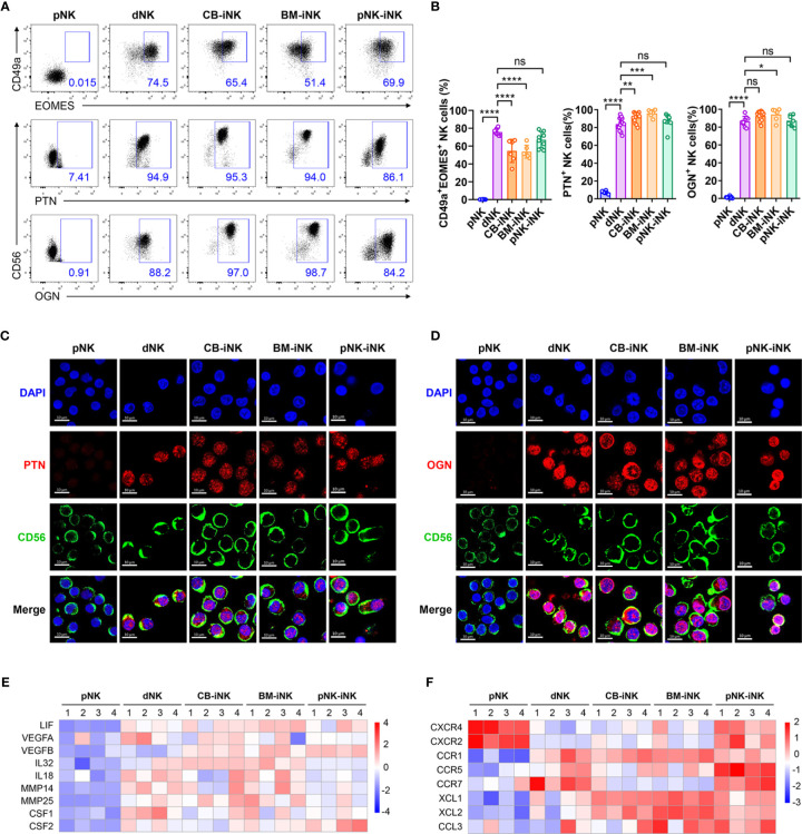
Figure 4.
The iNK cells have high expression of GPFs, proangiogenic factors, chemokines and chemokine receptors like dNK cells. (A, B) The percentage of CD49a+EOMES+ and GFP+ NK cells in each group was tested by flow cytometry. Representative density plots (A) and statistical calculation of all samples (B). Data represent means ± SD, n ≥ 6 in each group. Data were analyzed by one-way ANOVA. *p < 0.05; **p < 0.01; ***p < 0.005; ****p < 0.0001; ns, not significant. (C, D) CLSM images showing the secretion of PTN (C) and OGN (D) in pNKs, dNKs and other iNKs. Scale bars, 10 μm. (E) Some genes encoding the proangiogenic factors were selected for heat map analysis. (F) Some genes encoding the chemokines and chemokine-receptors were selected for heat map analysis.

