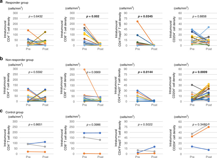
Fig. 3. Changes in intratumoral immune cells pre- and post-NAC treatment.
Relationship between the density of each immune cell type in the intratumoural area in pre-NAC tissue and post-NAC tissue in a responder, b non-responder, and c control groups. In responder and nonresponder groups, CD8+ T cells, and CD4+Foxp3+ T cells were significantly decreased in the post-NAC tissue compared with pre-NAC tissue. CD204+ cells were significantly increased in the post-NAC tissue compared with pre-NAC tissue in the non-responder group (p = 0.0009). In control group, each immune cells were not changed between pre-NAC tissue and post-NAC tissue.

