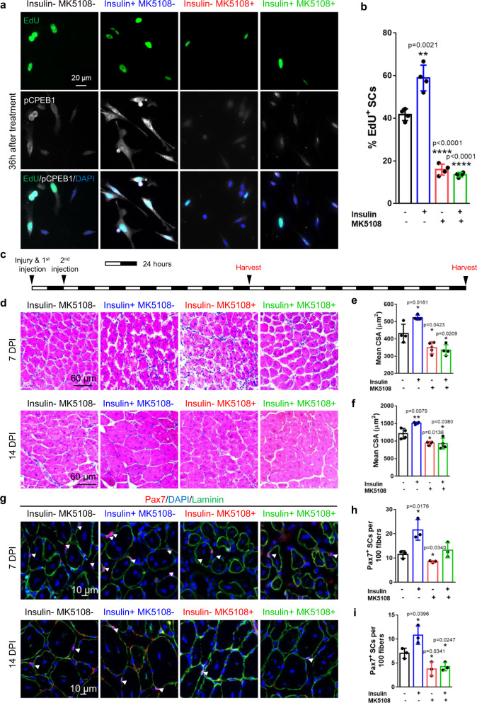
Fig. 7. Pharmacological manipulation of CPEB1 phosphorylation regulates SC activation, muscle regeneration and Pax7+ SC number after regeneration.
a, b FACS-isolated SCs were plated down and treated with MK5108 and/or insulin for 36 hours. Cells were also continuously supplied with EdU for EdU incorporation analysis. a 36 hours after MK5108 and/or insulin treatment, SCs were harvested for EdU detection and pCPEB1 immunostaining. Nuclei were stained with DAPI. b Quantification of EdU+ SCs after MK5108 and/or insulin treatment. (n = 4 independent experiments). c Timeline of muscle injury, intramuscular injection of MK5108 and/or insulin, and muscle regeneration study for d–i. d–f Tibialis anterior (TA) muscles were injured and injected with insulin and/or MK5108, then allowed to regenerate. d Histological analysis of the cross-sectioned TA muscles using hematoxylin and eosin (H&E) staining 7 days and 14-days post-injury (DPI). e, f Quantification of the size (in cross-sectional area) of regenerated fibers in d. (n = 4 independent experiments). g–i TA muscles were injured and injected with insulin and/or MK5108, then allowed to regenerate. g Immunostaining for Pax7 and laminin of cross-sectioned muscle fibers 7DPI and 14DPI. h, i Quantification of Pax7+ SCs in g. (n = 3 independent experiments). Data are presented as mean ± SD in panels b, e, f, h, i. The p values calculated by two-tailed unpaired t-test are used for comparing two groups in b, e, f, h, and i, ns not significant. Source data are provided as a Source Data file.

