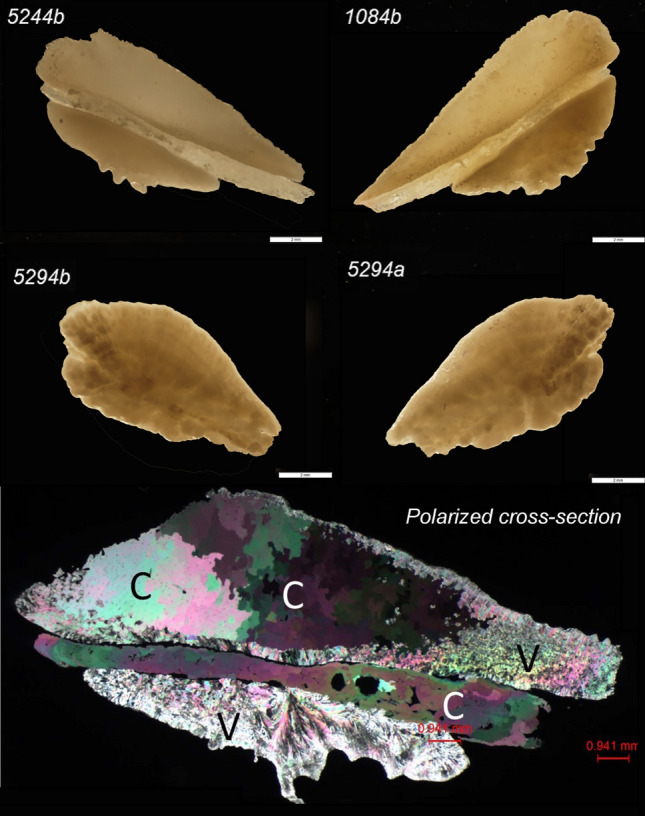
Figure 4.

Proximal (top) and distal (bottom) micrographs of Chinook salmon otoliths comprised of calcite and aragonite from fish 1084 and 5244 and two all aragonite otolith from fish 5294. The polarized cross-section panel shows a prepared (30 μm thick) thin section of 5244-b Chinook salmon otolith under cross-polarized light microscopy showing areas of calcite (C) and vaterite (V). The calcite is readily identified by its uniaxial (−) optic sign and large grain-sizes relative to vaterite.
