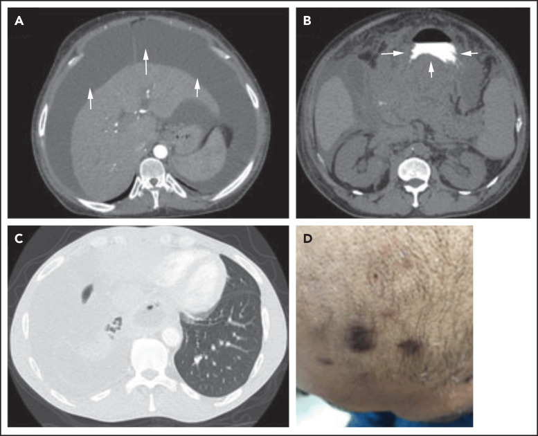
Figure 2.
Three different presentations of PEL. (A) Isolated malignant ascites in a patient with classic PEL (arrows). (B) EC/solid PEL infiltrating the stomach (arrows). Notice the mass displacing the oral contrast. (C-D) Isolated malignant right-sided pleural effusion (C) with concurrent KS (D). Notice the collapse of the right lung compared with the left (C), with characteristic purple KS lesions located on the side of the face in the same patient (D).

