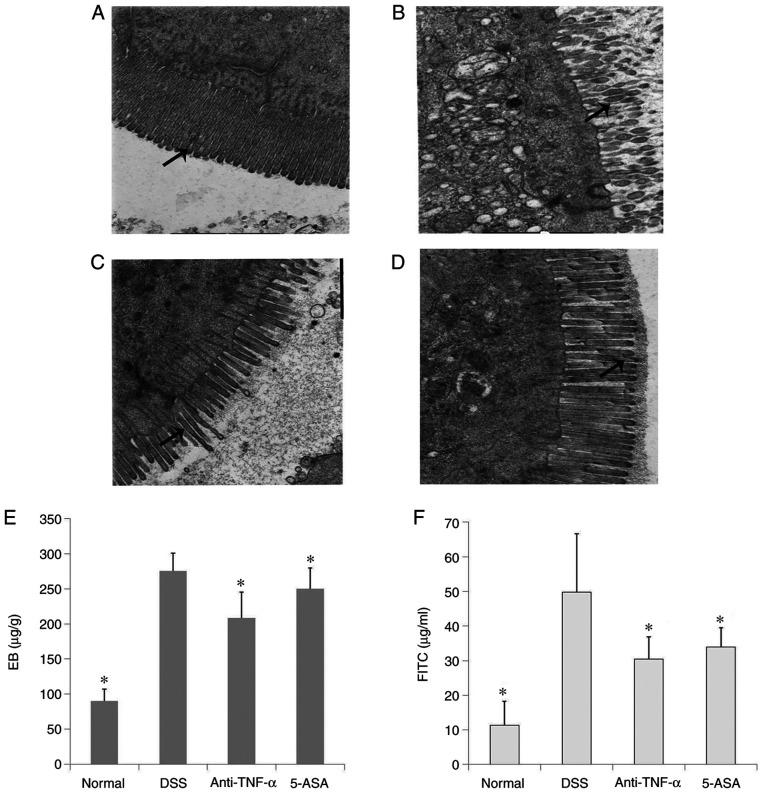
Figure 3.
Ultrastructure and function detection of the intestine. Ultrastructure of the intestinal mucosal barrier as observed by transmission electron microscopy in the (A) normal, (B) DSS, (C) anti-TNF-α and (D) 5-ASA groups. Magnification, ×20,000. Long-tailed arrows indicate surface microvilli. (E) The amount of EB permeating into the intestine isolated from mice in each of the four groups. Anti-TNF-α and 5-ASA decreased the level of intestinal EB staining in mice with DSS-induced colitis. (F) Plasma FITC-dextran 4000 level of the four groups. Anti-TNF-α and 5-ASA decreased the level of blood FITC in mice with DSS-induced colitis *P<0.05 vs. DSS. DSS, dextran sulfate sodium; EB, Evans blue; 5-ASA, 5-aminosalicyclic acid.

