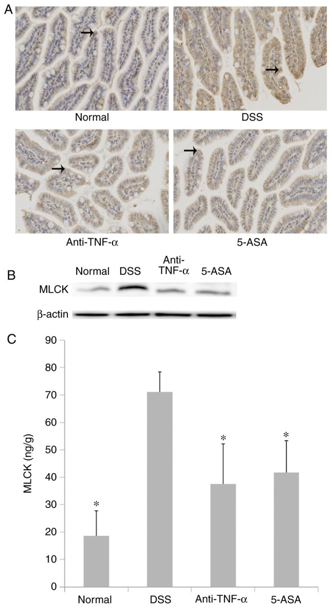
Figure 7.

Expression, distribution and activity of MLCK. (A) Expression and distribution of MLCK protein expression in the intestinal epithelium as observed via IHC. Anti-TNF-α and 5-ASA decreased the expression of MLCK protein in the intestinal epithelium of mice with DSS-induced colitis. Long-tailed arrows indicate MLCK expression. (B) The expression of MLCK in the intestinal epithelium as detected via western blotting. The results of western blotting were consistent with those in IHC. (C) MLCK enzymatic activity in the intestinal epithelium as detected by using ELISA. Anti-TNF-α and 5-ASA decreased MLCK enzymatic activity in the intestinal epithelium of mice with DSS-induced colitis. However, no significant difference was detected between anti-TNF-α and 5-ASA. *P<0.05 vs. DSS. MLCK, myosin light chain kinase; IHC, immunohistochemistry; DSS, dextran sulfate sodium; 5-ASA, 5-aminosalicyclic acid.
