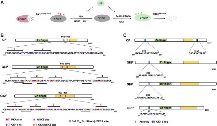
FIGURE 2.
Ci/Gli phosphorylation code. (A) Ci/Gli is phosphorylated on different sites by distinct sets of kinases depending on the Hh signaling states. In the absence of Hh, phosphorylation of CiF/GliF by PKA/CK1/GSK3 negatively regulates Hh signaling by converting CiF/GliF into CiR/GliR whereas in the presence of Hh, phosphorylation of CiF/GliF by Fu/Ulk3/Stk36 plays a positive role by promoting the formation of CiA/GliA. (B–C) Schematic representation of Ci and Gli showing the PKA/CK1/GSK3 phosphorylation sites (B) and Fu/Ulk3/Stk36 phosphorylation sites (C). Individual kinase sites are color coded. The underlines indicate the Slimb/β-TRCP binding sites in Ci/Gli. The green box represents the Zn-finger DNA-binding domain whereas the yellow box indicates the transactivation domain.

