. 2022 Feb 8;303(2):297–299. doi: 10.1148/radiol.213227

Figure 1:

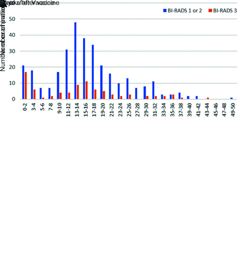
Graphs compare (A) patients with and without lymphadenopathy (LAD) at initial breast imaging after COVID-19 vaccination. Lymphadenopathy is seen most commonly in the first 2 weeks after vaccination, but can also persist at least 10 weeks. (B) Graphs compare patients with LAD and follow-up imaging. Bars show the percent of examinations assigned Breast Imaging Reporting and Data System (BI-RADS) category 1 or 2 (negative or benign findings) versus BI-RADS 3 (probably benign finding; short-term follow-up is recommended) recommendations by time after the vaccination. Twenty-five percent of examinations performed at 0–12 weeks were given BI-RADS 3 recommendations, and none of these patients were subsequently diagnosed with a new malignancy.

Graphs compare (A) patients with and without lymphadenopathy (LAD) at initial breast imaging after COVID-19 vaccination. Lymphadenopathy is seen most commonly in the first 2 weeks after vaccination, but can also persist at least 10 weeks. (B) Graphs compare patients with LAD and follow-up imaging. Bars show the percent of examinations assigned Breast Imaging Reporting and Data System (BI-RADS) category 1 or 2 (negative or benign findings) versus BI-RADS 3 (probably benign finding; short-term follow-up is recommended) recommendations by time after the vaccination. Twenty-five percent of examinations performed at 0–12 weeks were given BI-RADS 3 recommendations, and none of these patients were subsequently diagnosed with a new malignancy.