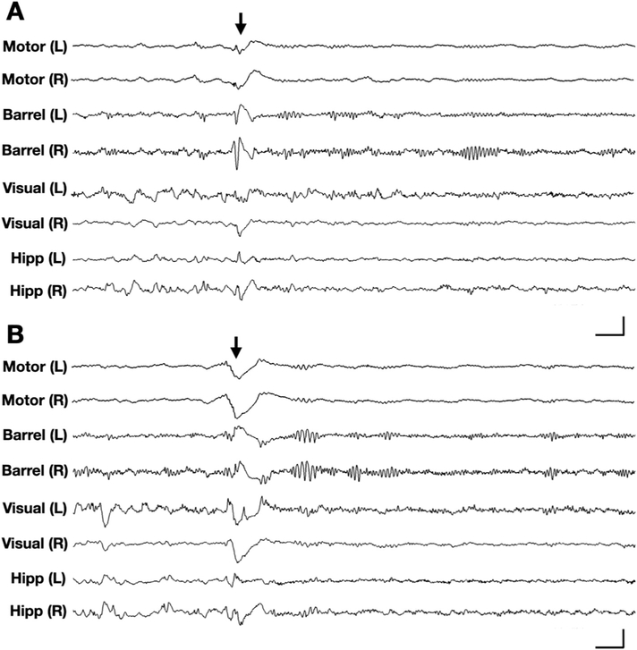
Fig. 2. Generalized epileptic spasms in Cdkl5KO/+ mice.
Representative 5-s traces of 8-channel intracranial EEG (channels: left/right motor cortex, barrel cortex, visual cortex, and hippocampus) recorded in a Cdkl5KO/+ mouse. (A) EEG corresponding to an epileptic spasm in a Cdkl5KO/+ mouse during sleep, characterized by bilaterally symmetric onset, spike-wave activity in barrel cortices and slow-wave activity in motor cortices. (B) EEG corresponding to another epileptic spasm in the same Cdkl5KO/+ mouse, several minutes later. This event is characterized by bilaterally symmetric onset, slow-wave activity in the motor and barrel cortices. (A-B) Vertical scale bars represent 1.8 mV, 1 mV, 1 mV, and 500 μV in bilateral motor cortex, barrel cortex, visual cortex, and hippocampal channels, respectively. Horizontal scale bars represent 0.25 s. Arrows indicate time of spasm onset.

