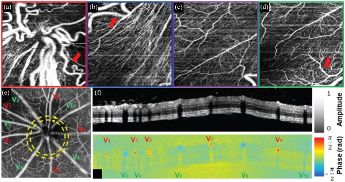
Figure 1.
(a)–(d) Handheld OCTA of ROP at the (a) optic nerve head, (b) peripapillary region, (c) perifoveal region, and (d) margin of the fovea. Visible-light OCT in a rodent model showing (e) OCTA projection with delineation of arteries (red) and veins (green), and circumpapillary (f) retinal structure and (g) Doppler blood flow cross section.
Source: Reprinted with permission from Viehland and colleagues 31 and Pi and colleagues. 43
OCT, optical coherence tomography; OCTA, optical coherence tomographic angiography; ROP, retinopathy of prematurity.

