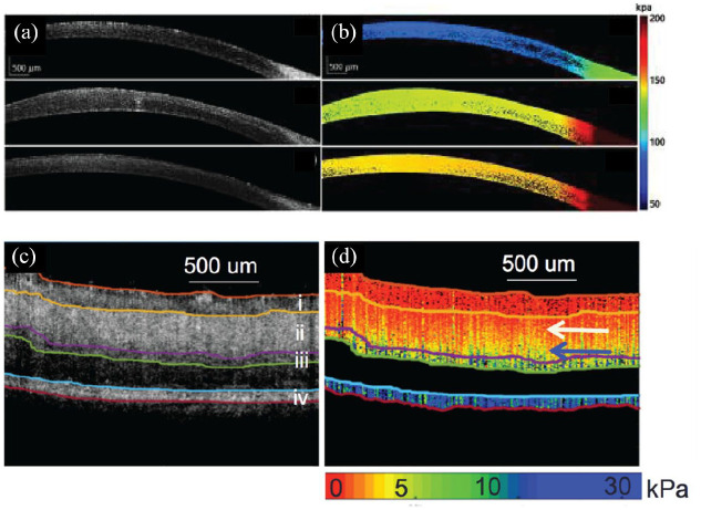
Figure 3.
OCE imaging of the (a), (b), cornea and (c), (d) retina. (a) Structural OCT and (b) OCE elastogram cross sections of in vivo rabbit cornea pre-, post-, and 1 week after CXL treatment (top to bottom, respectively). (c) Structural OCT and (d) OCE elastogram cross sections of ex vivo porcine retina showing differences in retinal layer stiffness.
Source. Reprinted with permission from Zhou and colleagues 127 and Qu and colleagues. 142
CXL, corneal collagen crosslinking; OCE, optical coherence elastography; OCT, optical coherence tomography.

