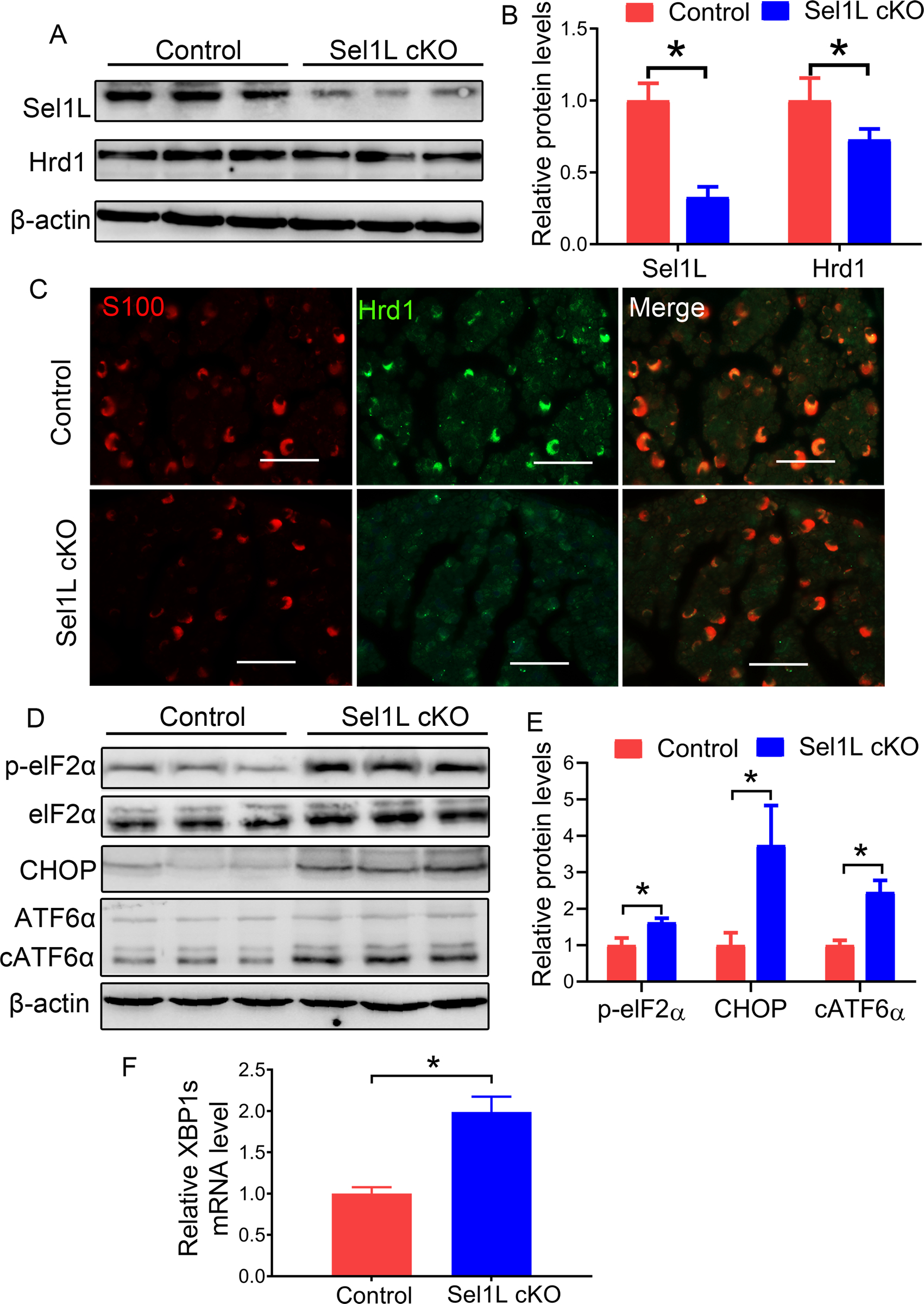
Figure 1. Sel1L deficiency led to elimination of Hrd1 and activation of the UPR in SCs.

A, B. Western blot analysis showed that the levels of Sel1L and Hrd1 were significantly decreased in the sciatic nerve of 3-w-old Sel1L cKO mice compared to control mice. C. S100 and Hrd1 double labeling revealed a dramatic reduction of Hrd1 immunoreactivity in SCs in the sciatic nerve of 3-w-old Sel1L cKO mice compared to control mice. D, E. Western blot analysis showed the elevated levels of p-eIF2α, CHOP, and cATF6α in the sciatic nerve of 3-w-old Sel1L cKO mice compared to control mice. F. Real time-PCR analysis revealed that the level of XBP1s mRNA was significantly increased in the sciatic nerve of 3-w-old Sel1L cKO mice compared to control mice. Scale bars: 50 μm. N = 4 animals. Statistical analyses were done with a t-test. Error bars represent SD, P < 0.05.
