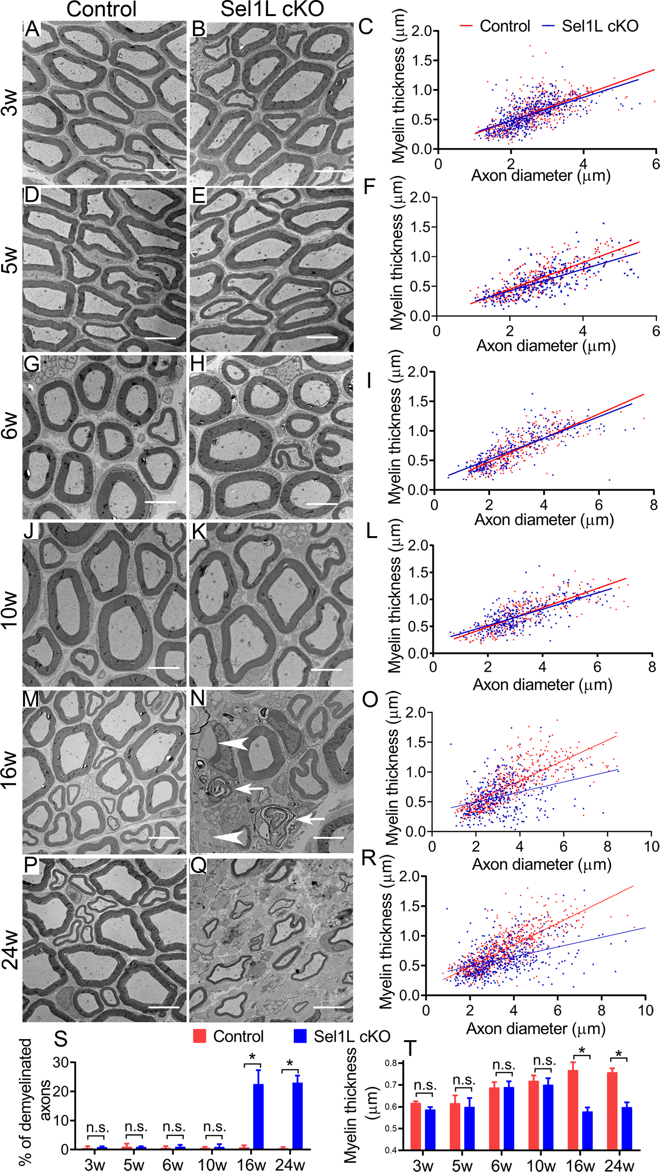
Figure 3. EM analysis revealed adult-onset demyelination in the PNS of Sel1L cKO mice.

A-L, S, T. EM analysis showed that the percentage of myelinated axons, the thickness of myelin, and the diameter of axons were comparable in the sciatic nerve of 3, 5, 6, 10-w-old Sel1L cKO mice and control mice. M-R, S, T. EM analysis showed that there were a significant increase in the percentage of demyelinated axons (arrowhead, naked axon; arrow, loose myelin) and a significant decrease in the thickness of myelin in the sciatic nerve of 16 and 24-w-old Sel1L cKO mice compared to control mice. Scale bars: 5 μm. N = 4 animals. Statistical analyses were done with a t-test. Error bars represent SD, n.s. not significant, P < 0.05.
