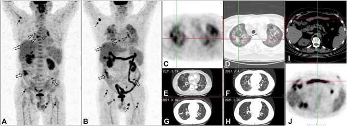
FIGURE 1.
A 58-year-old male patient with metastatic intrahepatic cholangiocarcinoma was treated with the third line of ipilimumab. After four cycles of treatment, compared with 18F-FDG PET/CT maximum intensity projection (MIP) image (A) before treatment, the posttreatment 18F-FDG PET/CT (B-D), (I,J) demonstrates the following: 1) multiple soft tissue density nodules in the peritoneum, which were larger in volume and higher in FDG uptake than before ((A,B) hollow arrows); 2) multiple nodules without FDG uptake in both lungs, some of which are larger in volume than before and some of which have no change compared with before (cannot be shown in MIP images- (A,B)); 3) the lymph nodes with increased FDG uptake in mediastinal areas and bilateral hilar, which were smaller in volume and FDG-uptake lower than before ((A,B) hollow triangles); and 4) multiple foci of increased FDG uptake in bones: the FDG-uptake degree in vertebral body of lumbar 4 was significantly lower than before ((A) gray triangle); the right acetabular lesion was the new lesion ((B) gray triangle); the remaining ostial lesions were higher than those before ((A,B) black line arrows). According to PERCIMT criteron: SMD was confirmed for the number of the newly FDG-positive lesions is less than 4. Meanwhile, in the light of imPERCIST5, PMD was also evaluated for the sum of SULpeak of the patient’s top five target lesions after treatment was more than 30% higher than that of the top five target lesions before treatment. As a result of clinical follow-up, the patient was confirmed PD and those lesion above were validated as metastases. PD was determined for the appearance of new lesion based on PECRIT. PMD was determined for new FDG-avid lesion at SCAN-2, which can be considered as UPMD in the line with iPERCIST. But the patient didn’t receive the next 18F-FDG PET/CT between days 21 and 28 after treatment or 4–8 weeks later, so it could not be evaluated according to PECRIT and iPERCIST. Follow-up results showed the PFS for ICI was 6 months. Additionally, this posttreatment PET/CT displayed: 1) newly patchy ground glass density foci in both lungs, with increased FDG uptake (C- PET axial image, D-CT axial image); 2) diffuse increased FDG uptake in ascending colon, transverse colon, descending colon, sigmoid colon and rectum (i-PET axial image) which is new compared with the previous one, and the corresponding colonic walls were not significantly thickened ((J)-CT axial image). The patchy ground glass shadows of both lungs gradually disappeared during chest CT follow-up ((E–H)-CT axial images). Combined with clinical information, the patchy ground glass shadows of both lungs were diagnosed as immunerelated pneumonia. Under the administration of MDT, the diffuse increase of glucose metabolism in the colon was considered as immune-related colitis.

