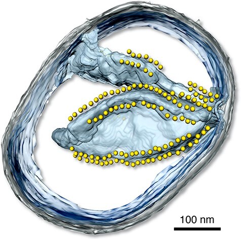
Fig. 5.

Three-dimensional tomographic volume of a small mitochondrion from P. anserina. The inner membrane is light blue, the outer membrane is grey. Yellow spheres indicate the catalytic F1 heads of ATP synthase dimers arranged in rows on the cristae ridges. Scale bar, 100 nm (B. Daum and W. Kühlbrandt, MPI of Biophysics, unpublished).
