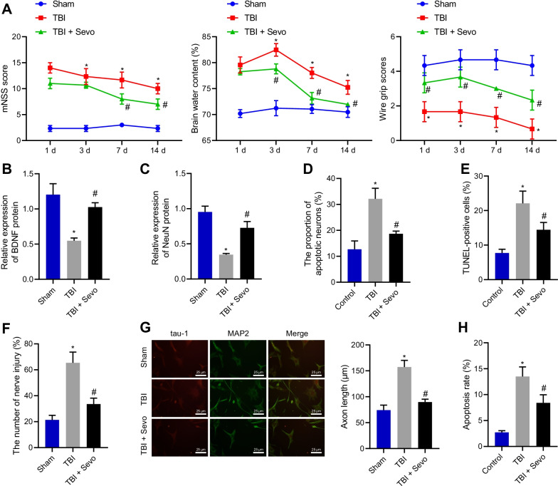
Fig. 1.
Sevo causes attenuation of neurological deficits and neuronal apoptosis in TBI rats. The sham-operated rats were utilized as controls, and TBI rats were treated or untreated with Sevo. A Neurological function evaluation by mNSS, brain water content measurement and motor function score in rats. B The protein expression of BDNF detected by Western blot analysis in the cortical tissue of rats. C Western blot analysis of the protein expression of NeuN in the cortical tissue of rats. D Nissl staining of the hippocampal neuronal damage in the cortical tissue of TBI rats. E TUNEL-positive cells in the cortical tissue of TBI rats. F Neuronal damage assessed by cell immunofluorescence assay (After primary hippocampal neurons were cultured for 48 h, Tau-1 was used to specifically identify axons, with red fluorescent markers in the figure; MAP2 was used to specifically identify dendrites, with green fluorescent markers, with a scale of 20 μm). G Axonal length measurement of hippocampal neurons. H The apoptosis of hippocampal neurons detected by flow cytometry. In panel A–E n = 12 for rats upon each treatment. *p < 0.05 vs. sham-operated rats or control cells; #p < 0.05 vs. TBI rats or cells. Cell experiments were conducted three times independently

