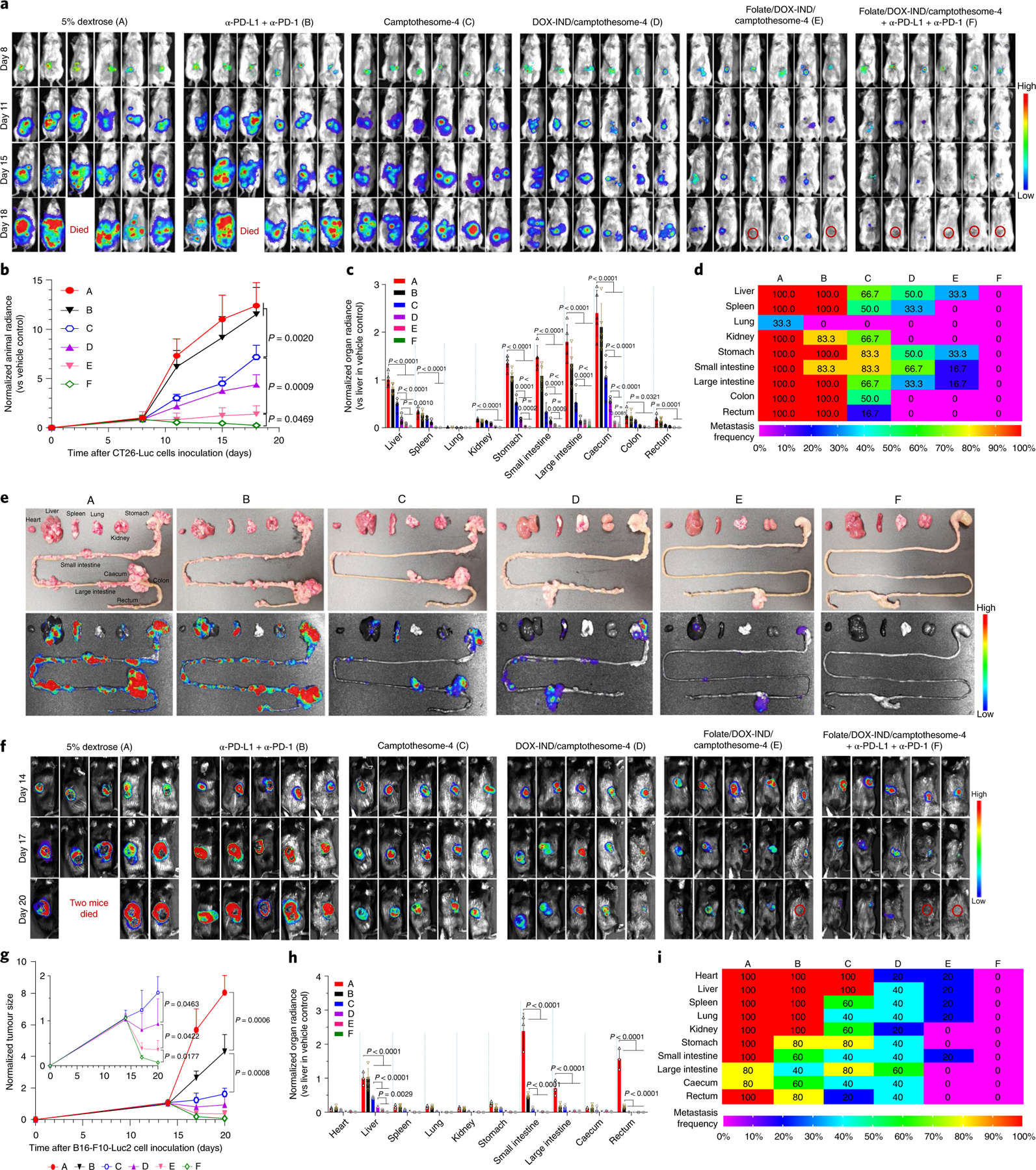
Fig. 6 |. Eradication of advanced and metastatic orthotopic CRC and subcutaneous melanoma tumours.

a–e, Therapeutic efficacy in orthotopic CRC tumour mouse model. Mice were inoculated with 2 × 10⁶ CT26-Luc cells (RPMI-1640/Matrigel, 3/1, v/v) into the caecum subserosa35. On day 8, mice (n = 6 mice; tumours, ~300 mg) were intravenously administered one dose of camptothesome-4, DOX-IND/camptothesome-4 or folate/DOX-IND/camptothesome-4 at the equivalent of 15 mg CPT kg−1 and 5 mg DOX-IND kg−1. α-PD-L1 and α-PD-1 were injected as described above. Lago imaging is shown for live mice (a). Red circles, tumour-free mice (one mouse each from the vehicle control and α-PD-L1 + α-PD-1 groups died on day 18). Quantitative bioluminescence intensity (QBI) for whole mouse tumour burden (b), QBI in various organs (c), a heatmap summarizing the tumour metastatic rate (d) and representative ex vivo photographs (e, upper panel) and bioluminescence imaging (e, lower panel) of various organs on day 18. f–i, Antitumour efficacy in melanoma-bearing C57BL/6 mice. Animals were subcutaneously inoculated with 0.1 × 10⁶ B16-F10-Luc2 cells. On day 14, mice (n = 5 mice; tumours, ~400 mm3) received the same treatments as in a. Lago imaging is shown for live mice (f). Red circle, tumour-free mouse. Two mice from the vehicle control group died on day 20. Normalized tumour size measured by a digital caliper (g), QBI in various organs (h) and a heatmap presenting the tumour metastatic rate (i). Data in b, c, g, h are expressed as mean ± s.d. Statistical significance was determined by one-way ANOVA followed by Tukey’s multiple comparisons test.
