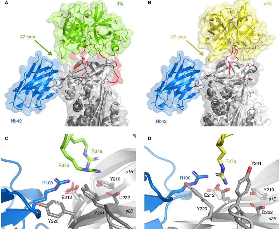
FIGURE 3.
Superimposition of the current crystal structure of PAI-1 (light gray) complexed with Nb42 (blue) and the previously determined structures of the Michaelis complexes between PAI-1 (dark gray) and tPA (green) (A, PDB: 5BRR), and PAI-1 (dark gray) and uPA (yellow) (B, PDB: 3PB1). The RCL of PAI-1 is red. C, Detail of the interaction interfaces of the PAI-1/Nb42 complex and the PAI-1/tPA complex. D, Detail of the interaction interfaces of the PAI-1/Nb42 complex and the PAI-1/uPA complex

