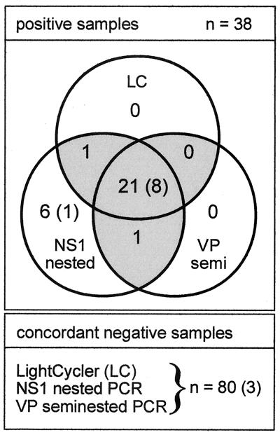
FIG. 3.
Detection of parvovirus B19 DNA in patient serum samples by three different DNA amplification methods. LC, LC-FRET assay specific for a 229-bp fragment of the NS-1 locus; VP, seminested PCR specific for a 174-bp fragment of the VP-1 locus; NS1, nested PCR specific for a 99-bp fragment of the NS-1 locus. Shaded areas of the Venn diagram indicate samples which were concordantly positive by at least two PCR formats. Numbers in parentheses indicate serum samples from an immunocompromised child; other samples originated from immunocompetent individuals.

