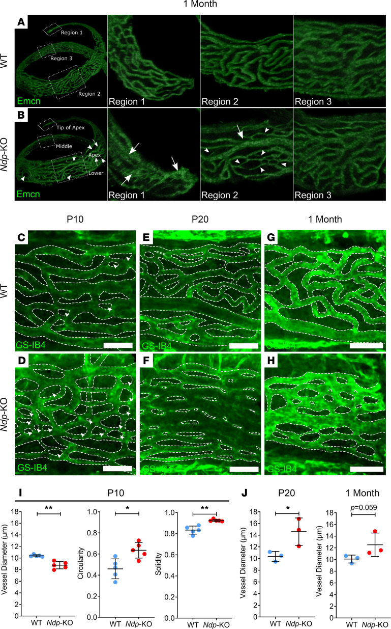
Figure 7. Postnatal onset of morphological abnormalities in stria vascularis capillaries in Ndp-KO.
The stria vascularis of 1-month-old mice was examined with an antibody targeting endomucin. (A and B) Panels display the middle-apical region of the stria vascularis cropped from a 3D image of whole cochleae from WT (A) and Ndp-KO (B) mice. The regions indicated by perforated boxes in A and B are displayed in adjacent panels. White arrows indicate dilated capillaries, white arrowheads indicate narrow capillaries and regions with sparse vessels. (C–H) The apical region of the stria vascularis was isolated and examined with GS-IB4. WT and Ndp-KO were examined at P10 (C and D), P20 (E and F), and 1 month (G and H). White arrowheads indicate intercapillary regions that are small and circular/oval in structure. (I) Quantification of vessel diameter and shape descriptor measurements of intercapillary regions for P10 stria vascularis; n = 5 WT, n = 5 Ndp-KO; bars indicate mean ± SD. (J) Quantification of vessel diameter in P20 and 1 month stria vascularis; n = 3 WT, n = 3 Ndp-KO; bars indicate mean ± SD. I (circularity) and J analyzed with an unpaired t test, I (vessel diameter and solidity) analyzed with Mann-Whitney test; *P < 0.05, **P < 0.01. Scale bars: 50 μm.

