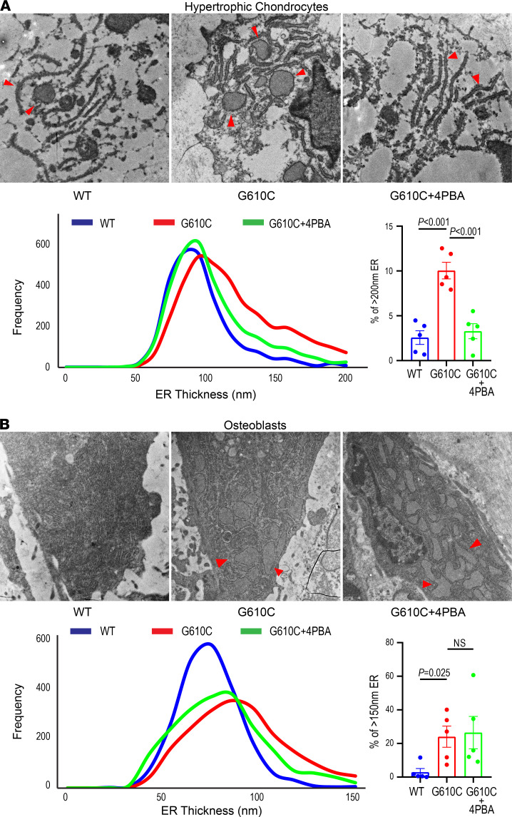
Figure 6. 4PBA treatment improves ER dilation in hypertrophic chondrocytes but to a lesser extent in osteoblasts.
(A and B) Representative transmission electron microscopic (EM) images of endoplasmic reticulum (ER, red arrowheads) in late hypertrophic chondrocytes from the tibial growth plate (A) and in tibial trabecular osteoblasts (B) after daily injections of PBS or 0.4 mg 4PBA in PBS (same animals as in Figures 4 and 5). Histograms show treatment effects on the ER thickness. Bar charts (mean ± SEM) show fractions of severely dilated ER in each mouse (≥ 200 nm thickness in HCs, ≥ 150 nm thickness in osteoblasts). ER morphology was examined by EM in n = 5 WT + PBS, n = 5 G610C + PBS, and n = 5 G610C + 4PBA animals from each of the groups selected based on their size being close to the mean value. We utilized the 2-tailed Student’s t test rather than 1-way ANOVA because meaningful statistical comparison could be performed only between WT + PBS and G610C + PBS animals (genotype effect) or between G610C + PBS and G610C + 4PBA animals (treatment effect), but not between WT + PBS and G610C + 4PBA animals.

