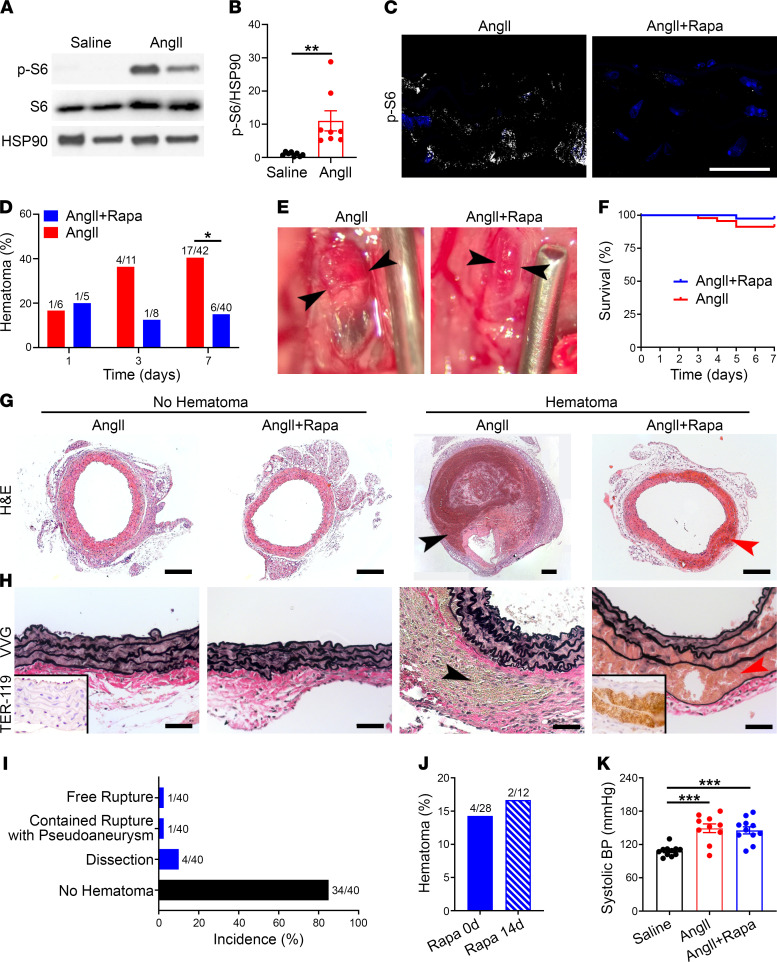
Figure 5. mTOR inhibition prevents aortic rupture and pseudoaneurysm but promotes dissection.
Apoe–/– mice were infused with AngII with or without rapamycin (Rapa) treatment for 7 days. (A) Western blot of aortas for phospho-S6 (p-S6) and S6. (B) Relative expression of phospho-S6 from pooled experiments (n = 8). (C) Confocal microscopy for phospho-S6 expression (white) and DAPI-labeled nuclei (blue) in media of suprarenal abdominal aortas, scale bar: 25 μm. (D) Incidence of aortic hematomas at 1–7 days. (E) Appearance of aortic hematomas (arrowheads), scale provided by needle of 1.27 mm outer diameter. (F) Survival of animals infused with AngII without (n = 42) or with (n = 40) rapamycin treatment for 7 days. (G) H&E stains showing medial thickening without hematomas compared with contained rupture (black arrowhead) and dissection (red arrowhead) in the absence or presence of rapamycin, respectively; scale bars: 200 μm (note different magnification of larger pseudoaneurysm). (H) Verhoeff-Van Gieson staining similarly shows thickened media without hematomas versus contained rupture (black arrowhead) and dissection (red arrowhead) in the absence or presence of rapamycin, respectively; TER-119 immunostain for erythrocytes in insets, scale bars: 50 μm. (I) Incidence of suprarenal abdominal aorta complications in AngII-infused Apoe–/– mice treated with rapamycin. (J) Incidence of aortic hematomas in AngII-infused mice treated with rapamycin from day 0 to 7 or from day –14 to 7. (K) Systolic blood pressure (BP) in Apoe–/– mice infused with saline (n = 11), AngII (n = 10), or AngII with rapamycin treatment (n = 11). Individual data shown for continuous variables, bars represent mean ± SEM, *P < 0.05, **P < 0.01, ***P < 0.001, unpaired, 2-tailed t test (B), Fisher’s exact test (D and J), and 1‑way ANOVA with Tukey’s multiple-comparison test (K).

