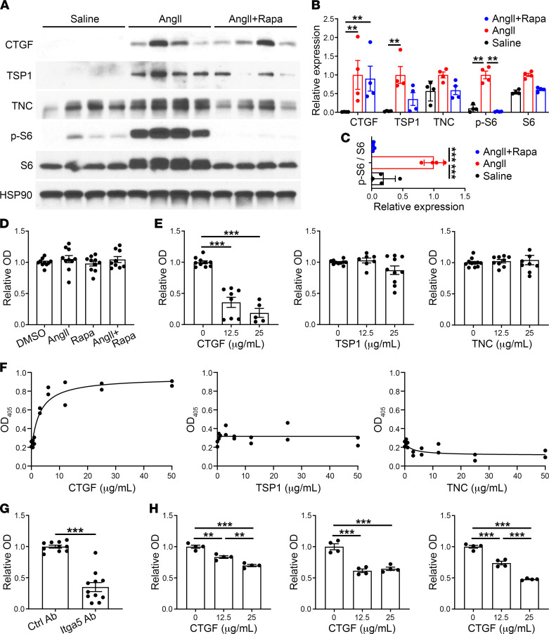
Figure 7. Rapamycin-insensitive CTGF inhibits SMC adhesion to exogenous ECM.
(A) Apoe–/– mice were infused with saline or AngII with or without rapamycin (Rapa) treatment for 7 days and the suprarenal abdominal aortas were analyzed by Western blot for CTGF, thrombospondin-1 (TSP1), tenascin-C (TNC), phospho-S6 (p-S6), and S6. (B) Densitometry of protein expression relative to HSP90 or (C) phospho-S6 expression relative to S6; expression normalized to peak levels with AngII treatment alone, (n = 4). (D) Colorimetric assay for number of murine aortic SMCs adherent to fibronectin-coated plates after 1 hour following cell pretreatment with vehicle, AngII at 100 nM, and/or rapamycin at 100 ng/mL for 45 minutes (n = 9–10, pooled from 3 experiments); OD405 readings normalized to vehicle-treated controls. (E) Similar fibronectin adhesion assay of SMCs pretreated with CTGF, thrombospondin-1, or tenascin-C at various doses for 45 minutes (n = 5–10, pooled from 3 experiments). (F) Colorimetric assay for number of SMCs adherent to plates coated with CTGF, thrombospondin-1, or tenascin-C at various concentrations (in the absence of fibronectin) after 1 hour (n = 2). (G) CTGF adhesion assay of SMCs pretreated with blocking antibody to integrin α5 (Itga5 Ab) or isotype-matched control antibody (Ctrl Ab) for 45 minutes (n = 10–11, pooled from 3 experiments). (H) Fibronectin adhesion assay of human aortic SMCs from 3 individuals pretreated with CTGF at various doses for 45 minutes (n = 4, shown separately for each subject). Individual data shown, bars represent mean ± SEM or lines represent nonlinear regression fitting by least-squares regression, **P < 0.01, ***P < 0.001, unpaired, 2-tailed t test (G), 1‑way ANOVA with Tukey’s multiple-comparison test (C–E and H), and 2‑way ANOVA with Sidak’s multiple-comparison test (B).

