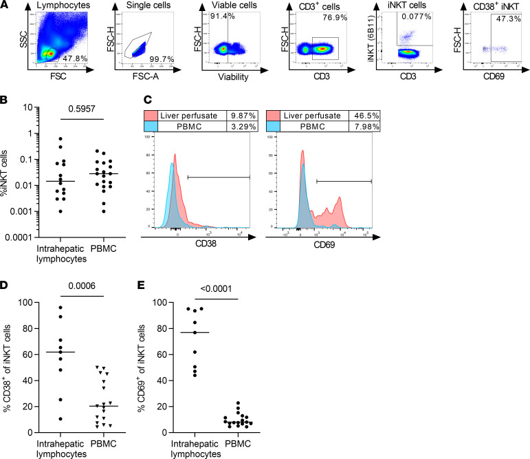
Figure 6. Intrahepatic iNKT cells are more activated than peripheral blood iNKT cells.
(A) Representative gating strategy for Vα24Jα18+ iNKT cells from liver perfusates. (B) Frequency of intrahepatic and peripheral blood iNKT cells of all CD3+ T cells from nonmatched donors (liver perfusate, n = 14; PBMC, n = 19; Mann-Whitney U test). (C) Expression of CD38 and CD69 on iNKT cells was analyzed in liver perfusates and peripheral blood of matched donors. A representative experiment is shown from 2 independent donors. (D and E) The frequency of CD38+ and CD69+ iNKT cells in liver perfusates and PBMCs from nonmatched healthy donors was analyzed by flow cytometry (liver perfusate, n = 9; PBMC, n = 17; unpaired t test).

