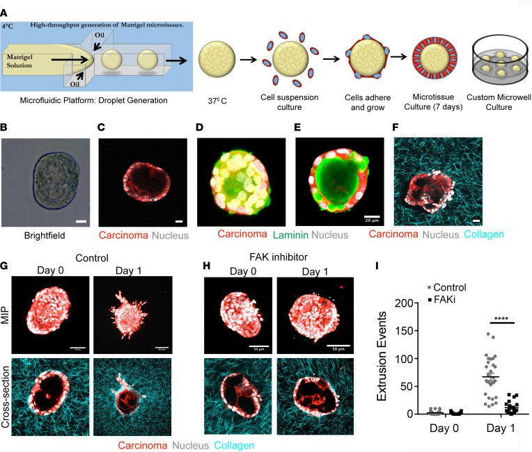
Figure 4. FAK inhibition abrogates collagen fiber–guided PDA cell extrusion in vitro.
(A) Schematic demonstrating our in vitro platform to mimic tumor cell extrusion from ductal structures into surrounding collagen. Matrigel droplets generated in a microfluidic device by orthogonal flow of Matrigel in an oil based medium, before coating them with primary KPCG or KPCT cells. Coated droplets are cultured individually in custom-fabricated microwells for several days before embedding in a collagen matrix. (B and C) Bright-field (B) and multiphoton excitation microscopy image (C) of a formed microtissue. (D and E) Maximum intensity projection (MIP) (D) and cross-sectional (E) MPE immunofluorescence micrographs show a typical droplet structure resembling the cross-section of a duct with basement membrane. (F) MPE/SHG imaging of a droplet embedded in collagen matrix. (G–I) Control (G) and FAK inhibitor (H) treated conditions, showing significant invasion in the control group at say 1, which is completely abrogated by inhibition of FAK, as quantified by single-cell extrusion analysis in (I); n = 20–30 droplets per condition from n = 2 experiments. Data are mean ± SEM; ****P < 0.0001 by ordinary 2-way ANOVA and Sidak’s multiple-comparison test. Scale bars: 20 μm (B–F) and 50 μm (G and H).

