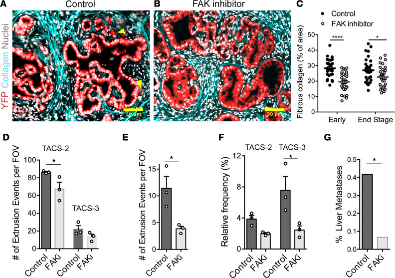
Figure 5. Inhibition of FAK alters frequency of collagen architectures, extrusion, and metastasis in PDA.
(A and B) Fluorescence micrographs of stained KPCY sections either treated with (A) vehicle control or (B) FAK inhibitor showing reduction in collagen deposition around ductal structures, smoother ductal boundaries, and reduction in cell extrusion; yellow arrowheads indicate single-cell extrusion events in the control sample. (C and D) Quantification of collagen content in KPC mice shows a reduction in deposition of fibrous collagen in both early (1.5 month) and end-stage mice, with a (D) concomitant reduction in TACS-like architectures in KPC mice. Note, the vast majority of PanIN lesions in FAKi-treated mice still retain TACS-2 and TACS-3 architectures in spite of lower overall collagen levels. (E) Number of extrusion events quantified from control and FAKi-treated mice showing reduced extrusion by FAK inhibition. (F) Extrusion events for the control and FAK inhibited groups quantified as a function of TACS-2 or TACS-3 architectures showing a decrease in extrusion and especially into TACS-3 regions. (G) Frequency of liver metastasis in control and FAKi-treated KPC mice showing reduction in liver metastases with FAK inhibitor treatment (data are from ref. 7). Data are mean — SEM (C–F), n = 29–36 FOV per group for C, n = 3 per group for D–F, and n = 6 for G. ****P < 0.0001, *P < 0.05 by 2-way ANOVA and Sidak’s multiple-comparison test for C, D, and F; *P < 0.05 for E by t test; *P < 0.05 by Fisher’s exact test for G. Scale bars: 50 μm.

