Figure 5.

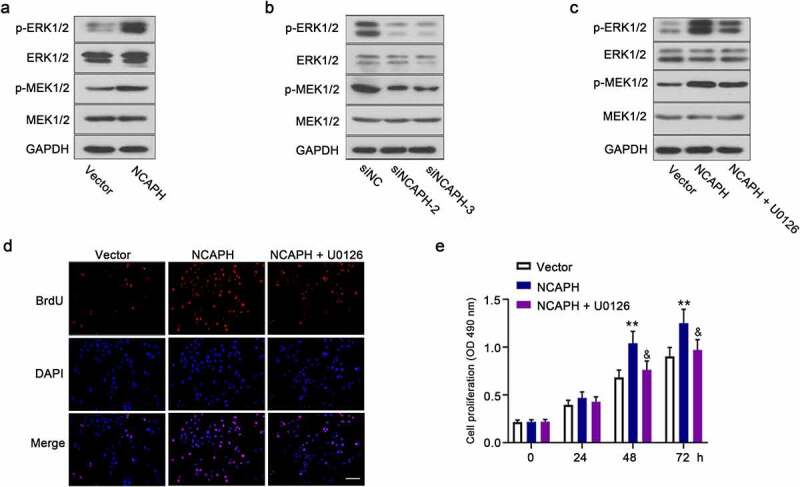
NCAPH promotes the activation of MEK/ERK signaling pathway in BC cells. (a,b) The protein levels of p-MEK1/2, MEK1/2, p-ERK1/2 and ERK1/2 in NCAPH overexpressing SW780 cells and NCAPH silenced UMUC3 cells were detected by Western blot. (c) SW780 cells were transfected NCAPH expression vector and treated with MEK1/2 inhibitor 10 μM U0126, the protein levels of p-MEK1/2, MEK1/2, p-ERK1/2 and ERK1/2 were detected by Western blot. (d,e) The cell proliferation of BC cells was determined by BrdU assay (Scale bar, 100 μm) and MTT assay. The data were presented as the mean ± SD. Vector versus NCAPH, **P < 0.01; NCAPH versus NCAPH + U0126, &P < 0.05. No significant difference between Vector and NCAPH, NCAPH and NCAPH + U01260 group at 0 and 24 h.
