Figure 6.

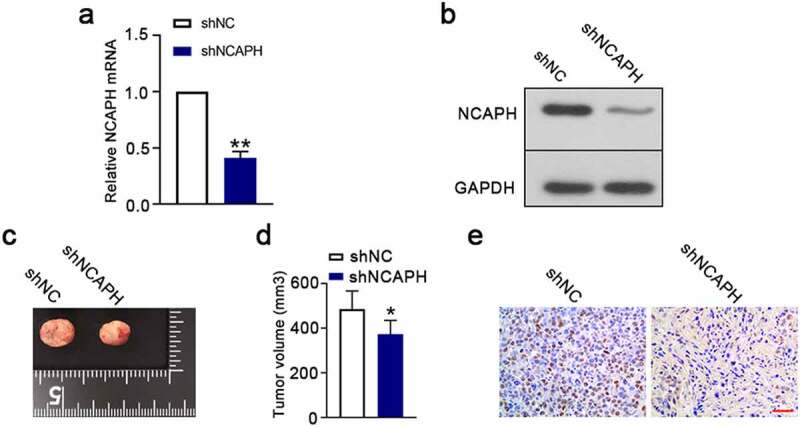
Knockdown of NCAPH suppresses tumor growth in vivo. (a,b) A total of 5 × 10⁶ shRNA-mediated NCAPH silenced UMUC3 cells were subcutaneously injected into nude mice. The tumor tissues were immediately harvested at 25 days post implantation. The mRNA and protein levels of NCAPH in tumor tissues were determined by qRT‐PCR and Western blot. (c,d) Tumor volume was evaluated at day 25. (e) The expression of Ki67 (brown staining) in tumor tissues was detected using immunohistochemistry staining. Scale bar, 50 μm. The data were presented as the mean ± SD. **P < 0.01, *P < 0.05.
